. 2002 Jun 11:2:11.
doi: 10.1186/1472-6750-2-11.

Embryonic stem cells and mice expressing different GFP variants for multiple non-invasive reporter usage within a single animal

Affiliations

Embryonic stem cells and mice expressing different GFP variants for multiple non-invasive reporter usage within a single animal

Anna-Katerina Hadjantonakis et al. BMC Biotechnol. .

Abstract

Background: Non-invasive autofluorescent reporters have revolutionized lineage labeling in an array of different organisms. In recent years green fluorescent protein (GFP) from the bioluminescent jellyfish Aequoria Victoria has gained popularity in mouse transgenic and gene targeting regimes 1. It offers several advantages over conventional gene-based reporters, such as lacZ and alkaline phosphatase, in that its visualization does not require a chromogenic substrate and can be realized in vivo. We have previously demonstrated the utility and developmental neutrality of enhanced green fluorescent protein (EGFP) in embryonic stem (ES) cells and mice 2.

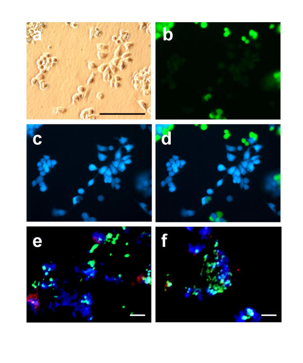
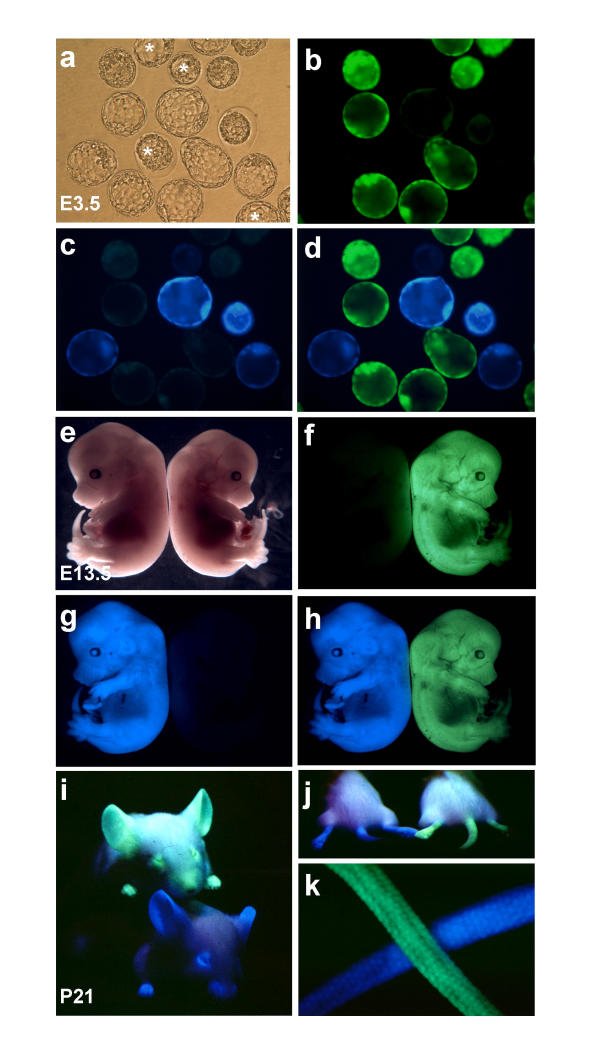
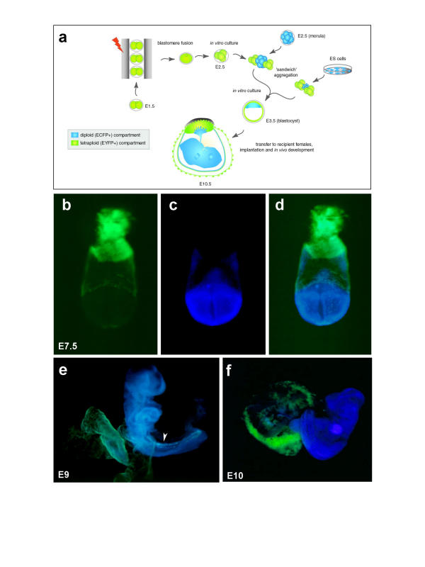
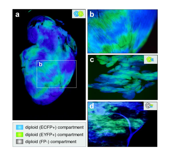
Results: In this study we have used embryonic stem (ES) cell-mediated transgenesis to test the enhanced cyan fluorescent protein (ECFP) and enhanced yellow fluorescent protein (EYFP), two mutant and spectrally distinct color variants of wild type (wt) GFP. We have also tested DsRed1, the novel red fluorescent protein reporter recently cloned from the Discostoma coral by virtue of its homology to GFP. To this end, we have established lines of ES cells together with viable and fertile mice having widespread expression of either the ECFP or EYFP GFP-variant reporters. However, we were unable to generate equivalent DsRed1 lines, suggesting that DsRed1 is not developmentally neutral or that transgene expression cannot be sustained constitutively. Balanced (diploid <-> diploid) and polarized (tetraploid <-> diploid) chimeras comprising combinations of the ECFP and EYFP ES cells and/or embryos, demonstrate that populations of cells expressing each individual reporter can be distinguished within a single animal.

Conclusions: GFP variant reporters are unique in allowing non-invasive multi-spectral visualization in live samples. The ECFP and EYFP-expressing transgenic ES cells and mice that we have generated provide sources of cells and tissues for combinatorial, double-tagged recombination experiments, chimeras or transplantations.


Figures

Figure 1
Figure 1
Non-invasive multiple reporter visualization in ES cells. (a-d) a mixture of CK6/ECFP (ECFP+) and YC5/EYFP (EYFP+) ES cells at low density. (a) image taken under bright field with no epifluorescence. (b) dark field image taken through an EYFP filter. (c) dark field image taken through an ECFP filter. (d) dark field double exposure image acquired by consecutively using ECFP and EYFP filters. ECFP+ cells can clearly be discerned from EYFP+ cells. (e-f) culture of mixed populations of ES cells comprising ECFP+, EYFP+ and RFP+ cells. Scale bars represent 100 μm.
Figure 2
Figure 2
Dual non-invasive reporter visualization in embryos and adult mice. Transgenic animals are hemizygous for either the CK6/ECFP or YC5/EYFP transgenes. (a-d) blastocyst stage (E3.5) embryos, that are either fluorescent or non-fluorescent. Fluorescent embryos are either blue/cyan or yellow/green, thereby being hemizygous for either the CK6/ECFP or YC5/EYFP transgenes respectively. Non-fluorescent embryos (marked with an asterix on panel a) are of the wild type outbred ICR strain. (a) image taken under bright field with no epifluorescence. (b) dark field image taken through an EYFP filter. (c) dark field image taken through an ECFP filter. (d) dark field double exposure image acquired by consecutively using ECFP and EYFP filters. (e-h) two transgenic (TG/+) E13.5 embryos dissected free of their extraembryonic membranes, one is blue/cyan fluorescent (CK6/ECFP transgenic, left) and the other is yellow/green fluorescent (7YC5/EYFP transgenic, right). (e) image taken under bright field with no epifluorescence. (f) dark field image taken through an EYFP filter. (g), dark field image taken through an ECFP filter. (h) double exposure acquired by consecutively using ECFP and EYFP filters. (i-k) two three week old transgenic mice each exclusively expressing either the ECFP or EYFP reporter. All images shown are double exposures taken by consecutively using ECFP and then the EYFP filters under epifluorescence. (i) CK6/ECFP (TG/+) transgenic mouse bottom, YC5/EYFP (TG/+) transgenic mouse bottom. (j) CK6/ECFP (TG/+) transgenic mouse left, YC5/EYFP (TG/+) transgenic mouse right. (k) higher maginification view of the tails of the mice in i and j.
Figure 3
Figure 3
Dual non-invasive reporter visualization in chimeric embryos generated by tetraploid <-> diploid aggregation. In all panels the tetraploid compartment is yellow/green fluorescent (EYFP+) and the diploid is blue/cyan fluorescent (ECFP+). (a) schematic representation of the tetraploid procedure. Electrofusion of E1.5 embryos to render the blastomeres tetraploid, in vitro culture, and subsequent aggregation schemes using double-tagged compartments. Sandwich aggregations were made using either ECFP+ (diploid) morulae or ES cells. (b-d) anterior view of an E7.5 double-tagged polarized chimera dissected free of its deciduum and therefore missing primary giant cells and parietal endoderm. Here the epiblast (lower half of embryo) and its derivatives are ECFP+ whereas the extraembryonic ectoderm, the visceral endoderm and trophoblast are EYFP+ (upper half or embryo). (b) dark field image taken through an EYFP filter. (c) dark field image taken through an ECFP filter. (d) dark field double exposure image acquired by consecutively using ECFP and EYFP filters. (e) dark field double filter exposure of an E9 chimera dissected free of all extraembryonic membranes except for a small piece of yolk sac (lower left). The embryo itself is ECFP+ whereas the yolk sac both ECFP+ and EYFP+ in the mesoderm and endoderm respectively. A small number of EYFP+ cells are also observed in the ventral midline of the embryo (arrowhead). This observation has been noted previously in tetraploid chimeras, and is presumed to represent a small population of visceral endoderm cells that fail to be displaced by the definitive endoderm at earlier stages. (f) dark field double filter exposure of an E10 chimera. The embryo is exclusively ECFP+ whereas its placenta (to the left of the embryo) is predominantly EYFP+. The labyrinthine trophoblast layer of the placenta comprises both ECFP+ and EYFP+ cells.
Figure 4
Figure 4
Dual non-invasive reporter visualization in the organs of adult chimeras. All images depict dark field double exposures acquired by consecutively using ECFP and EYFP filters under dark field epifluorescence. Details of aggregates are schematized at the top right of each panel. (a-c) heart and pancreas from double-tagged double-compartment adult chimeras. (a) view of the surface of an adult heart. (b) close-up surface view of ventricle of a chimeric heart generated by aggregation of two diploid morulae, one hemizygous for the CK6/ECFP (ECFP+) transgene and the other hemizygous for the YC5/EYFP (EYFP+) transgene. The regions of yellow/green and cyan fluorescence are clearly mutually exclusive. The striations in the fields of fluorescence represent the proliferative zones present within the ventricle. (c) chimeric pancreas generated by aggregation of a morula hemizygous for the YC5/EYFP transgene with CK6/ECFP ES cells. The restricted fields of fluorescence represent the proliferative zones present within the pancreas. (d) liver from a double-tagged triple-compartment adult chimera. A non-fluorescent morula was aggregated with two clumps of ES cells, one carrying the CK6/ECFP transgene (ECFP+) and the other carrying the YC5/EYFP transgene (EYFP+). An ECFP+ vessel has clonally proliferated and infiltrated the liver lobe (top). Note that unlike in heart and pancreas there appears to be more interspersed blue/cyan vs green/yellow fluorescence present in the liver, possibly reflecting greater cell intermingling during the genesis of this organ.

Similar articles

Cited by

References

    1. Chalfie M, Tu Y, Euskirchen G, Ward WW, Prasher DC. Green fluorescent protein as a marker for gene expression. Science. 1994;263:802–5. - PubMed
    1. Hadjantonakis AK, Gertsenstein M, Ikawa M, Okabe M, Nagy A. Generating green fluorescent mice by germline transmission of green fluorescent ES cells. Mech Dev. 1998;76:79–90. doi: 10.1016/S0925-4773(98)00093-8. - DOI - PubMed
    1. Chalfie M. Green fluorescent protein. Photochem Photobiol. 1995;62:651–6. - PubMed
    1. Cormack BP, Valdivia RH, Falkow S. FACS-optimized mutants of the green fluorescent protein (GFP). Gene. 1996;173:33–8. doi: 10.1016/0378-1119(95)00685-0. - DOI - PubMed
    1. Zernicka-Goetz M, Pines J, McLean Hunter S, Dixon JP, Siemering KR, Haseloff J, Evans MJ. Following cell fate in the living mouse embryo. Development. 1997;124:1133–7. - PubMed

Publication types

Substances