. 2002 Jun 15;16(12):1466-71.
doi: 10.1101/gad.991402.

FIH-1 is an asparaginyl hydroxylase enzyme that regulates the transcriptional activity of hypoxia-inducible factor

Affiliations

FIH-1 is an asparaginyl hydroxylase enzyme that regulates the transcriptional activity of hypoxia-inducible factor

David Lando et al. Genes Dev. .

Abstract

Mammalian cells adapt to hypoxic conditions through a transcriptional response pathway mediated by the hypoxia-inducible factor, HIF. HIF transcriptional activity is suppressed under normoxic conditions by hydroxylation of an asparagine residue within its C-terminal transactivation domain, blocking association with coactivators. Here we show that the protein FIH-1, previously shown to interact with HIF, is an asparaginyl hydroxylase. Like known hydroxylase enzymes, FIH-1 is an Fe(II)-dependent enzyme that uses molecular O(2) to modify its substrate. Together with the recently discovered prolyl hydroxylases that regulate HIF stability, this class of oxygen-dependent enzymes comprises critical regulatory components of the hypoxic response pathway.


Figures

Figure 1
Figure 1
Predicted secondary structure of FIH-1. A multiple sequence alignment was generated using Jpred (Cuff et al. 1998) for amino acid residues 179–300 of human FIH-1. Related proteins are indicated by their gi accession numbers. Segments predicted to be in an extended conformation (β-strand) are denoted by arrows. Identical residues are shaded in black, and similar residues are shaded in gray. Asterisks indicate critical amino acids for Fe(II)-binding in characterized hydroxylase enzymes.
Figure 2
Figure 2
Effect of FIH-1 on HIF-1α and HIF-2α transactivation domain activity. (A) 293T cells were cotransfected with expression vectors for either the Gal4 DNA binding domain (GalDBD) or the indicated GalDBD/HIF-1α 727–826 or GalDBD/HIF-2α 774–874 chimeras, a Gal4 response element-driven luciferase reporter, and wild-type FIH-1 (wt) or H199A or D201A substitution mutants as indicated. (B) 293T cells were cotransfected with either GalDBD/HIF-1α 727–826 or Gal/HIF-2α 774–874 chimeras, a Gal4 response element-driven luciferase reporter, and 200 ng of wild-type FIH-1 plasmid. Transfected cells were subjected to increasing (0–200 μM) dimethyl-oxalylglycine (DMOG) treatment. Data are the average of three transfections +/− S.D.
Figure 3
Figure 3
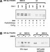
In vitro hydroxylation of HIF-2α 774–874 by FIH-1 inhibits interaction with p300. 35S-labeled HIF-2α 774–874 wild-type (wt) or the N851A mutant were treated with MBP-FIH-1 (wt or D201A mutant) in the presence of Fe(II), ascorbate (asc), and 2-oxoglutarate (2OG) or the hydroxylase inhibitor DMOG, then incubated with immobilized GST–p300 CH1. 35S-labeled HIF-2α 774–874 bound to the GST–p300 CH1 domain was visualized following SDS-polyacrylamide gel electrophoresis. FIH-1 activity is inhibited by 1 mM CoCl2 (A) or 2 mM DMOG (B).
Figure 4
Figure 4
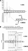
HIF-2α 774–874 is efficiently hydroxylated at residue Asn 851 by FIH-1 in vitro. (A) MALDI-TOF-MS spectra of trypsin-digested Trx6H HIF-2α 774–874 after treatment with FIH-1 in the presence of 2-oxoglutarate (2OG, upper spectrum) or DMOG (lower spectrum). The tryptic peptide YDCEVNVPVPGSSTLLQGR (846–864) is hydroxylated (+16 daltons) after treatment in the presence of 2OG, but not DMOG (magnified below). (B) Tandem MS sequencing of the hydroxylated and nonhydroxylated HIF-2α 846–864 tryptic peptides after FIH-1 treatment with 2-oxoglutarate (2OG) or DMOG, respectively, shows the hydroxylated residue is Asn 851. For instance, b- and y-type fragment ions were observed with the 2090.9 unmodified peptide that covered nearly the entire peptide sequence (lower spectrum). The modified 2106.9 peptide produced some fragment ions (upper spectrum) at the same m/z values as the unmodified peptide. These coincidental fragments correspond to portions of the sequence that did not contain Asn 851. Fragments were also observed for the modified sequence at m/z values of +16 daltons more than the Asn 851-containing fragments of the unmodified sequence (indicated by b+ and y+ in the upper panel). The boundary of the coincident and +16-dalton fragment ions is indicated by the symbol N+ on the upper panel.
Figure 5
Figure 5
Effect of hypoxia on FIH-1 inhibition of transactivation domain activity. Human embryonic kidney 293 cells were transfected with a Gal4-tk-luc reporter DNA, the HIF-1α 727–826/Gal4 chimera (wild-type or the N803A substitution mutant) and FIH-1 (wild-type or the D201A substitution mutant) or a LacZ control. Cells were incubated for 19 h under normoxic conditions or for 5 h under normoxic conditions followed by 14 h under hypoxic conditions (0.5% O2). FIH-1, but not the D201A mutant, suppresses CAD activation under hypoxic (0.5% O2) conditions. FIH-1 suppression of CAD activity under normoxic conditions is not observed under these conditions as baseline luciferase activity levels are at the level of detection. Mutation of the asparagine to alanine renders the CAD insensitive to either FIH-1 or oxygen concentration.

References

    1. Aravind L, Koonin EV. The DNA-repair protein AlkB, EGL-9, and leprecan define new families of 2-oxoglutarate- and iron-dependent dioxygenases. Genome Biol. 2001;2:1–8. - PMC - PubMed
    1. Bruick RK, McKnight SL. A conserved family of prolyl-4-hydroxylases that modify HIF. Science. 2001;294:1337–1340. - PubMed
    1. Carrero P, Okamato K, Coumailleau P, O'Brien S, Tanaka H, Poellinger L. Redox-regulated recruitment of the transcriptional coactivators CREB-binding protein and SRC-1 to hypoxia-inducible factor 1α. Mol Cell Biol. 2000;20:402–415. - PMC - PubMed
    1. Clifton IJ, Hsueh L-C, Baldwin JE, Harlos K, Schofield CJ. Structure of proline 3-hydroxylase. Evolution of the family of 2-oxoglutarate dependent oxygenases. Eur J Biochem. 2001;268:6625–6636. - PubMed
    1. Cockman ME, Masson N, Mole DR, Jaakkola P, Chang GW, Clifford SC, Maher ER, Pugh CW, Ratcliffe PJ, Maxwell PH. Hypoxia inducible factor-α binding and ubiquitylation by the von Hippel-Lindau tumor suppressor protein. J Biol Chem. 2000;275:25733–25741. - PubMed

Publication types

MeSH terms