. 2002 Jun 15;16(12):1488-97.
doi: 10.1101/gad.985002.

Neurogenin 3 is essential for the proper specification of gastric enteroendocrine cells and the maintenance of gastric epithelial cell identity

Affiliations

Neurogenin 3 is essential for the proper specification of gastric enteroendocrine cells and the maintenance of gastric epithelial cell identity

Catherine S Lee et al. Genes Dev. .

Abstract

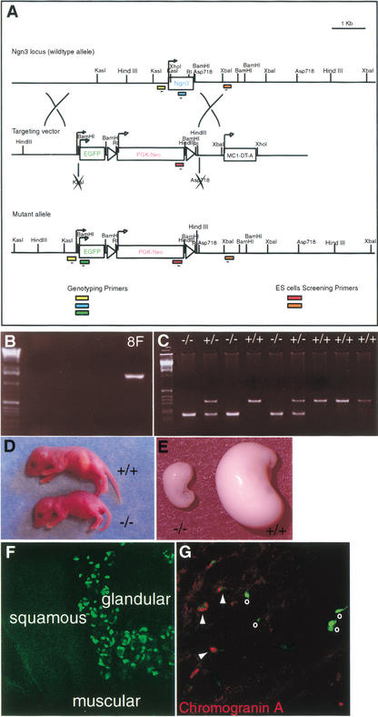
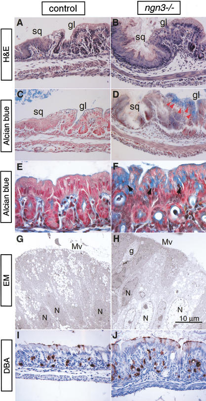
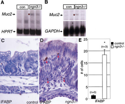
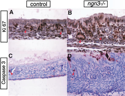
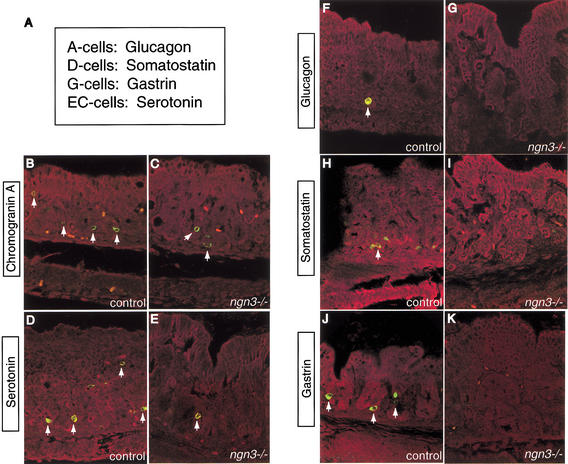
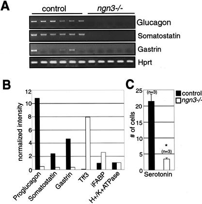
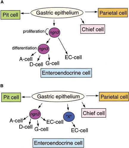
The notch signaling pathway is essential for the endocrine cell fate in various tissues including the enteroendocrine system of the gastrointestinal tract. Enteroendocrine cells are one of the four major cell types found in the gastric epithelium of the glandular stomach. To understand the molecular basis of enteroendocrine cell development, we have used gene targeting in mouse embryonic stem cells to derive an EGFP-marked null allele of the bHLH transcription factor, neurogenin 3 (ngn3). In ngn3(-/-) mice, glucagon secreting A-cells, somatostatin secreting D-cells, and gastrin secreting G-cells are absent from the epithelium of the glandular stomach, whereas the number of serotonin-expressing enterochromaffin (EC) cells is decreased dramatically. In addition, ngn3(-/-) mice display intestinal metaplasia of the gastric epithelium. Thus, ngn3 is required for the differentiation of enteroendocrine cells in the stomach and the maintenance of gastric epithelial cell identity.


Figures

Figure 1
Figure 1
Targeting strategy for ngn3 inactivation. (A) Gene structure of the ngn3 locus (top). Targeting vector used for homologous recombination in ES cells (middle). Gene structure of the targeted allele (bottom). Not all of the restriction enzymes are depicted in the diagram. (B) An ES clone (8F) that has undergone homologous recombination was identified using ES screening primers (indicated by red and orange boxes in A). (C) Genotyping by PCR of a litter from an intercross of heterozygous mice. Three sets of genotyping primers (indicated by yellow, blue, and green boxes in A) were used to generate the wild-type (295 bp) and mutant (188 bp) allele bands. (D) Photograph of 3-day-old wild-type (top) and ngn3−/− mice (bottom) on a mixed 129Sv × C57BL6 background. Ngn3−/− animals were smaller, dehydrated, and diabetic. (E) Photograph of the stomachs of 3-day-old wild-type (right) and ngn3−/− mice (left). Note the smaller size of the ngn3−/− stomach. (F) Confocal images of EGFP expression in the P3 heterozygous animal. ngn3–EGFP expression is detected only in the glandular stomach. (G) ngn3–EGFP is expressed in the enteroendocrine precursor cells. Confocal image of heterozygous stomach stained using a Cy3-conjugated secondary antibody against Chromogranin A that allows simultaneous detection of Chromogranin A (red) and ngn3 expression (green). Some cells expressed EGFP alone (indicated by o), whereas others were colabeled with both EGFP and Chromogranin A (indicated by arrowheads).
Figure 2
Figure 2
Abnormal stomach development in ngn3−/− mice. Paraffin sections of stomachs obtained from 3-day-old control (A,C,E,,I) and ngn3−/− mice (B,D,F,J) were stained with histological and lectin stains. Hematoxylin and eosin staining of control (A) and ngn3−/− (B) stomachs. Note increased diameter and disorganized appearance of the gastric epithelium in ngn3−/− mice. (C,E) Alcian blue, which detects acid mucins, normally does not stain the gastric epithelium, but detects ectopic acid mucins in the ngn3−/− mice (D,F, indicated by red and black arrowheads). Electron micrographs illustrate the ultrastructural features of the stomach. (G) An image of a control stomach showing normal epithelial morphology, whereas goblet-like cells were present in the ngn3−/− stomach (H). Control and ngn3−/− glandular epithelium were stained with Dolichos biflorus (DBA) lectin, which labels parietal cells, with horseradish peroxidase substrate (brown). The DBA staining shows no difference in the number and distribution of parietal cells between the control (I) and ngn3−/− gastric epithelium (J). Magnification for A–D and I–J is 400×, E–F is 600×, and G–H is 5000×. (sq) Squamous epithelium; (gl) glandular epithelium; (g) goblet cell; (Mv) microvilli; (N) nucleus.
Figure 3
Figure 3
Up-regulation of intestine-specific gene expression in the stomach of ngn3−/− mice. Muc2 mRNA–PCR and RNase protection assay (RPA) analysis. (A) RT–PCR analysis of total RNA isolated from whole stomach of control and ngn3−/− mice. Muc2 expression (215 bp) is detected in the ngn3−/− stomach (n = 2), whereas no Muc2 expression is found in the control (n = 2). Hypoxanthine phosphoribosyl transferase (HPRT) was used as a loading control (130 bp). (B) RPA analysis of 10 μg of total RNA from 3-day-old control (n = 2) and ngn3−/− stomach (n = 2). Glyceraldehyde phosphate dehydrogenase (GAPDH) served as loading control. Muc2 mRNA was again seen in ngn3−/− stomach. Notably, the ngn3−/− stomach that showed higher expression of Muc2 by RT–PCR analysis also showed higher expression of Muc2 by RPA (indicated by dot in both A and B). (C–D) Immunohistochemical detection of intestinal fatty acid-binding protein (iFABP) in ngn3−/− stomach. Paraffin sections from 3-day-old control and ngn3−/− glandular stomach were stained with an antiserum specific to iFABP (indicated by red arrowheads). iFABP is expressed by the misspecified gastric epithelial cells in ngn3−/− mice (D), but was not detected in controls (C). Magnification, 400×. (E) iFABP-positive cells were counted from control (n = 3) and ngn3−/− stomachs (n = 3). Y-axis represents the iFABP-positive cell counts. (*) P < 0.02 by two-tailed Student's t-test.
Figure 4
Figure 4
Ngn3−/− gastric mucosa does not display abnormal cell proliferation or programmed cell death. Proliferating cells stained by Ki67 are found in the gastric glands (arrowheads), mesenchymal, and muscular layers of control (A) and ngn3−/− stomach (B). There was no significant difference in the number or localization of proliferating cells between the mutant and control groups. Caspase 3 immunostaining (arrowheads) showed no significant changes in numbers of apoptotic cells between control (C) and ngn3−/− (D stomachs). Magnification, 400×.
Figure 5
Figure 5
Ngn3 is required for the differentiation of enteroendocrine cells in the gastric epithelium. (A) Classification of enteroendocrine cells by their main secretory products. Immunofluorescence was performed on paraffin sections from 2–3-day-old control (B,D,F,H,J) and ngn3−/− (C,E,G,I,K) glandular stomach. Immunostained cells are labeled in green for the enteroendocrine cell-specific antigen indicated at left (labeled by white arrows), and images were captured by confocal microscopy. Evan's blue was used as counterstain for the tissues and is visualized in red in all sections (Beaulieu 1997). (B) Chromogranin A, a general endocrine cell maker, labels all subtypes of enteroendocrine cells in the gastric epithelium of control mice. (C) The number of Chromogranin A-expressing cells is reduced in the ngn3−/− gastric epithelium. (D) An enterochromaffin (EC) cell-specific marker, serotonin, is normally expressed by the EC-cells in the control stomach. (E) The number of serotonin-positive cells is reduced in the ngn3−/− stomach. (F) An A-cell-specific enteroendocrine cell marker, glucagon, is normally present in the gastric epithelium of control stomach. (G) No glucagon-positive cells are found in the ngn3−/− gastric epithelium. (H) Somatostatin, a D-cell-specific enteroendocrine cell maker, is found in the control gastric epithelium. (I) Somatostatin expression is not present in the ngn3−/− stomach. (J) Gastrin expression is found in the G-cells of control gastric epithelium. (K) No gastrin-positive cells are found in the ngn3−/− gastric epithelium. Magnification, 400×.
Figure 6
Figure 6
Analysis of mRNA levels of secretory products of enteroendocrine cells and gastrointestinal marker genes. (A) Glucagon, Somatostatin, and Gastrin mRNA are absent in 2–3-day-old ngn3−/− stomachs. RT–PCR analysis was performed using the primers indicated and the products separated on 1.5% ethidium bromide-stained agarose gels. Hprt was used as an internal control. n = 6 for controls and n = 5 for ngn3−/− stomachs. (B) Summary of genes with altered expression levels identified by microarray analysis. Y-axis shows the normalized intensity levels and X-axis shows the various genes examined. n = 2 for controls and n = 3 for ngn3−/− stomachs. (C) Quantification of serotonin-positive cells. Ten sections from each stomach were counted for both control and ngn3−/− animals. n = 3 for both control (black bar) and ngn3−/− stomachs (white bar). Y-axis represents the serotonin-positive cell counts. Serotonin-positive cells were decreased sixfold in the ngn3−/− stomachs. (*) P < 0.02 by two-tailed Student's t-test.
Figure 7
Figure 7
Two models for the proposed role of ngn3 during enteroendocrine cell differentiation in the gastric epithelium. (A) Ngn3 is needed for the proliferation of all enteroendocrine cells, but only required for the differentiation of A-, D-, and G-cells. EC-cells can differentiate in an ngn3-independent pathway, but their number is reduced in ngn3−/−; mice as the total pool of enteroendocrine precursor cells is reduced. (B) ngn3 is absolutely required for the differentiation of A-, D-, and G-cells. However, differentiation of the EC-cells can occur from either the ngn3 lineage or another pathway regulated by an unidentified factor X.

Similar articles

Cited by

References

    1. Artavanis-Tsakonas S, Rand MD, Lake RJ. Notch signaling: Cell fate control and signal integration in development. Science. 1999;284:770–776. - PubMed
    1. Beaulieu JF. Extracellular matrix components and integrins in relationship to human intestinal epithelial cell differentiation. Prog Histochem Cytochem. 1997;31:1–78. - PubMed
    1. Borges M, Linnoila RI, van de Velde HJ, Chen H, Nelkin BD, Mabry M, Baylin SB, Ball DW. An achaete-scute homologue essential for neuroendocrine differentiation in the lung. Nature. 1997;386:852–855. - PubMed
    1. Duncan SA, Nagy A, Chan W. Murine gastrulation requires HNF-4 regulated gene expression in the visceral endoderm: Tetraploid rescue of Hnf-4(−/−) embryos. Development. 1997;124:279–287. - PubMed
    1. Falk P, Roth KA, Gordon JI. Lectins are sensitive tools for defining the differentiation programs of mouse gut epithelial cell lineages. Am J Physiol. 1994;266:G987–G1003. - PubMed

Publication types

MeSH terms