. 2002 Jun 15;16(12):1498-508.
doi: 10.1101/gad.989102.

Characterization of HCP-6, a C. elegans protein required to prevent chromosome twisting and merotelic attachment

Affiliations

Characterization of HCP-6, a C. elegans protein required to prevent chromosome twisting and merotelic attachment

Jeffrey H Stear et al. Genes Dev. .

Abstract

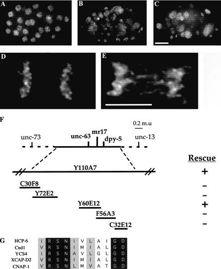
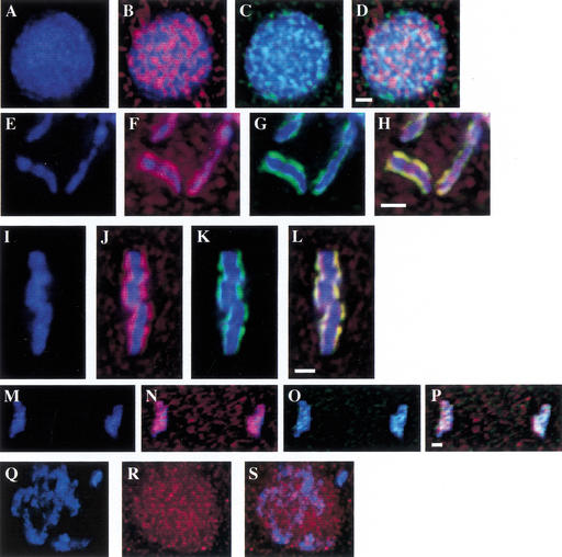
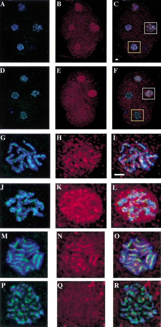
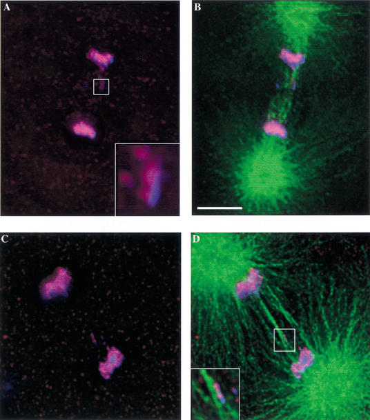
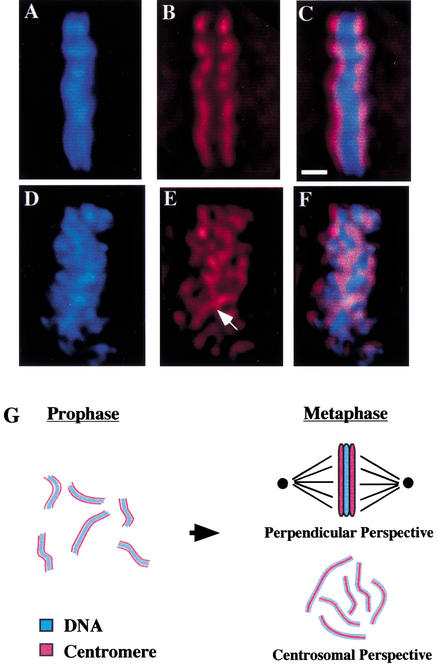
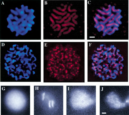
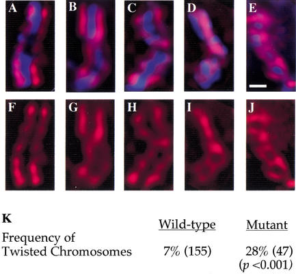
Previous studies of mitosis show that capture of single kinetochores by microtubules from both centrosomes (merotelic orientation) is a major cause of aneuploidy. We have characterized hcp-6, a temperature-sensitive chromosome segregation mutant in C. elegans that exhibits chromosomes attached to both poles via a single sister kinetochore. We demonstrate that the primary defect in this mutant is a failure to fully condense chromosomes during prophase. Although centromere formation and sister centromere resolution remain unaffected in hcp-6, the chromosomes lack the rigidity of wild-type chromosomes and twist around the long axis of the chromosome. As such, they are unable to establish a proper orientation at prometaphase, allowing individual kinetochores to be captured by microtubules from both poles. We therefore propose that chromosome rigidity plays an essential role in maintaining chromosome orientation to prevent merotelic capture.


Figures

Figure 1
Figure 1
hcp-6 embryos contain aneuploid nuclei, suggesting a defect in mitotic chromosome segregation. Embryos were isolated from adults and stained with DAPI to visualize DNA. (A) Note the uniform size and distribution of the nuclei in a wild-type embryo. (B) In hcp-6 embryos, the size and intensity of the DAPI staining vary greatly, indicating that DNA content is not consistent between individual cells. Furthermore, the nuclei are unevenly dispersed throughout the embryo, indicating multinucleate blastomeres. (C) RNAi of Y110A7A.1 generates a chromosome segregation phenotype identical to that of hcp-6. (D,E) Anaphase is disrupted in hcp-6 embryos. One-cell embryos undergoing anaphase were examined. (D) In the wild type, note the clean separation of sister chromatids. (E) In contrast, observe the strings of DNA suspended between the sister chromatids in hcp-6, indicative of anaphase bridging. (F) Cloning of HCP-6. Genetic and physical maps of the region surrounding hcp-6. Two YACs (Y110A7 and Y60E12) were able to rescue the embryo lethal phenotype associated with hcp-6. (G) Alignment of HCP-6 with XCAP-D2-like proteins from S. pombe (T43519), S. cerevisiae (S51408), X. laevis (AAC64359), and H. sapiens (NP055680) (GenBank accession numbers in parentheses). Bar, 5 μm.
Figure 2
Figure 2
Localization of HCP-6 throughout the cell cycle. (AP) Wild-type embryos showing DNA (blue), anti-HCP-6 (red), or anti-HCP-3 (green) staining, or merged images. Colocalization between HCP-6 and HCP-3 is shown as yellow. (AD) At interphase, this antigen is visible as punctate dots distributed throughout the nucleus. (EH) In prophase nuclei, HCP-6 forms two parallel lines on individual prophase chromosomes, a pattern suggesting centromere localization. (IL) At metaphase, the HCP-6 protein is present on the poleward faces of the metaphase plate. (MP) During anaphase, HCP-6 remains associated with the separating groups of sister chromatids. (QS) hcp-3(RNAi) embryos stained with anti-HCP-6 (red). In the absence of HCP-3, HCP-6 does not assemble onto mitotic chromosomes properly. Bars, 1 μm.
Figure 3
Figure 3
HCP-6 fails to localize to mitotic chromosomes in hcp-6 mutants. Following a 1-h shift to 26°C, embryos were stained with anti-HCP-6 (red) and anti-HCP-3 (green). DNA was visualized with DAPI (blue). Four-cell embryos at the same stage of development were examined. The AB blastomeres (bottom and left) are in metaphase. The remaining two blastomeres are in prophase, with the EMS blastomere (right) at a slightly later point in mitosis. (AC) In wild-type cells, as was described above, HCP-6 can be seen associating with both prophase and metaphase chromosomes. (DF) In contrast, although the HCP-6 antigen is present in prophase nuclei, it is completely absent from hcp-6 metaphase chromosomes. (GR) Magnified views of the EMS prophase nuclei (white boxes, GL) and the AB metaphase plates (yellow boxes, MR). In wild-type cells (GI), the HCP-6 protein can be seen associating with prophase chromosomes. However, in hcp-6 (JL), this antigen is distributed throughout the nucleus, and shows no enrichment at the chromosomes. Inspection of wild-type metaphase plates (MO) demonstrates that HCP-6 is easily detectable at the centromere of metaphase chromosomes. In contrast, although HCP-3 localizes to the centromere of mutant chromosomes (PR), no HCP-6 staining is observed. Bars, 1 μm.
Figure 4
Figure 4
hcp-6 chromosomes display merotelic orientation. hcp-6 embryos were stained with anti-HCP-3 (red), anti-tubulin (green), and DAPI (blue) following a 2-h shift to 23°C. Images of one-cell embryos undergoing anaphase were then collected. (B, D, and D, inset) The tubulin staining demonstrates that microtubules from both poles are associating with the lagging chromosomes in hcp-6 embryos. A higher-resolution image of the lagging chromosome (A, inset) reveals that only one line of HCP-3 is present, suggesting that this DNA body represents a single chromatid. Taken together, these observations indicate that merotelic chromosomes are generated in the hcp-6 background. Bar, 5 μm.
Figure 5
Figure 5
Centromere organization is disrupted in hcp-6. All data were collected from one-cell embryos following a 1-h shift to 26°C and show DNA (blue), anti-HCP-3 (red), or a merged image of both. (AF) HCP-6 chromosomes are not organized properly within the metaphase plate (perpendicular perspective). (AC) In wild-type cells, HCP-3 localizes to the poleward faces of the metaphase plate, as described above. (DF) In hcp-6 embryos, although linear aggregates of HCP-3 staining, corresponding to the centromeres of individual chromosomes are visible at metaphase, their localization is not restricted to the poleward faces of the metaphase plate. Instead, they run throughout the width of the metaphase plate and associate with both poles simultaneously (see arrow). (G) At metaphase, all of the chromosomes have aligned themselves onto the metaphase plate, a disc-like structure which lies in the middle of the cell. When viewed from the “perpendicular perspective,” (top right) such that the poles lie to the left and right of the image, the chromosomes in the metaphase plates merge together and appear as a solid bar of DNA. The centromeres, which have maintained their 180° of opposition, are oriented towards the poles, and lie on either side of the DNA. It is also possible to view a metaphase plate from the “centrosomal perspective” (bottom right) such that the metaphase plate lies flat on the plane of the page, and the centrosomes lie above and below the page. In this case, one is able to look down at the metaphase plate and individual sister centromeres, each lying on top of its corresponding metaphase chromosome. If one were able to view further down through the page, the opposing set of sister centromeres would then become visible. Bar, 1 μm.
Figure 6
Figure 6
hcp-6 possesses a chromosome condensation defect. (AF) Metaphase plates in AB blastomeres (4-cell embryo) stained with anti-HCP-3 (red) and DAPI (blue) and viewed from the centrosomal perspective. (AC) In wild-type cells, 12 fully compact chromosomal masses are evident, each with a corresponding centromere that runs the length of the chromosome. (DF) The chromosomes in an hcp-6 metaphase plate are longer and thinner, indicating a failure to fully condense. Although aggregates of HCP-3 are visible along the chromosomes, uninterrupted stretches of centromeric staining cannot be detected. (GJ) hcp-6 chromosomes display a prophase condensation defect. The images shown here represent GFP∷ histone-labeled nuclei at interphase (M,O) and prophase (N,P). (N) Note the fully condensed chromosomes that are detectable in the wild type. (P) In hcp-6, although some degree of condensation is evident, the chromosomes have not achieved a wild-type level of compaction. Bars, 1 μm.
Figure 7
Figure 7
hcp-6 chromosomes are less rigid than wild-type. (AJ) Prophase nuclei from one-cell embryos were examined in both wild-type and hcp-6 backgrounds. Embryos were stained with DAPI to visualize DNA (blue) and anti-HCP-3 (red). Images of prophase chromosomes stained with both DAPI and anti-HCP-3 (AE) or anti-HCP-3 alone (FJ). In both wild-type and hcp-6 embryos, the majority of prophase chromosomes displayed two parallel lines of HCP-3 staining which ran the length of the chromosome (A,F). More often in hcp-6, but also in the wild type, chromosomes displaying single 180° twists were observed (B,C,G,H). In hcp-6 embryos, chromosomes displaying two (D,I) or more (E,J) twists were also observed, patterns which were not seen in the wild type. The chromosomes displayed in (A–C,F–H) were obtained from wild-type embryos; (D,E,I,J) were collected from hcp-6 embryos. (K) Quantitative analysis of the incidence of twisted chromosomes. The number of chromosomes examined is in parentheses. The frequency of twisted chromosomes was increased fourfold in hcp-6 embryos. Bars, 1 μm.

Similar articles

Cited by

References

    1. Albertson DG, Thomson JN. The kinetochores of Caenorhabditis elegans. Chromosoma. 1982;86:409–428. - PubMed
    1. Bhalla N, Biggins S, Murray AW. Mutation of YCS4, a budding yeast condensin subunit, affects mitotic and nonmitotic chromosome behavior. Mol Biol Cell. 2002;13:632–645. - PMC - PubMed
    1. Brenner S. The genetics of Caenorhabditis elegans. Genetics. 1974;77:71–94. - PMC - PubMed
    1. Buchwitz BJ, Ahmad K, Moore LL, Roth MB, Henikoff S. A histone-H3-like protein in C. elegans. Nature. 1999;401:547–548. - PubMed
    1. Cimini D, Fioravanti D, Salmon ED, Degrassi F. Merotelic kinetochore orientation versus chromosome mono-orientation in the origin of lagging chromosomes in human primary cells. J Cell Sci. 2002;115:507–515. - PubMed

Publication types

MeSH terms