. 2002 Jun 15;16(12):1568-81.
doi: 10.1101/gad.986602.

Targeted mutagenesis by homologous recombination in D. melanogaster

Affiliations

Targeted mutagenesis by homologous recombination in D. melanogaster

Yikang S Rong et al. Genes Dev. .

Abstract

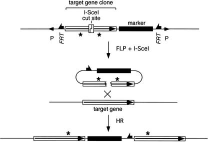
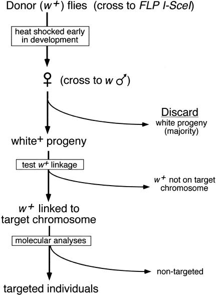
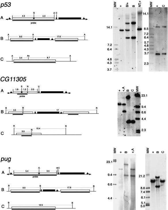
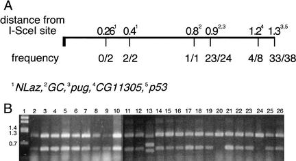
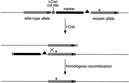
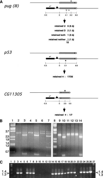
We used a recently developed method to produce mutant alleles of five endogenous Drosophila genes, including the homolog of the p53 tumor suppressor. Transgenic expression of the FLP site-specific recombinase and the I-SceI endonuclease generates extrachromosomal linear DNA molecules in vivo. These molecules undergo homologous recombination with the corresponding chromosomal locus to generate targeted alterations of the host genome. The results address several questions about the general utility of this technique. We show that genes not near telomeres can be efficiently targeted; that no knowledge of the mutant phenotype is needed for targeting; and that insertional mutations and allelic substitutions can be easily produced.


Figures

Figure 1
Figure 1
General form of ends-in gene targeting. The recombinogenic donor molecule is generated by FLP and I-SceI action on a P-element donor construct, causing excision of the target-homologous sequence and the marker gene, and cutting at the I-SceI site. HR with the chromosomal target locus generates a tandem duplication of the sequence that was present in the donor with incorporation of the marker gene between duplicated target segments. Mutations engineered into the donor (indicated as asterisks) may be carried into both copies of the target gene.
Figure 2
Figure 2
Schematic representation of the targeting process. In the first step, donor-bearing flies are crossed to flies carrying the heat-inducible FLP and I-SceI genes. The progeny of this cross are heat-shocked during the first few days of their development, and the eclosing daughters are crossed to w males. Because FLP-mediated excision is very efficient (typically >99% with the 38°C, 1-h heat shock used here; Golic and Golic 1996), most progeny have white eyes. The w+ progeny are screened to look for movement of the w+ marker from the donor chromosome to the target chromosome using test crosses with marked chromosomes (Rong and Golic 2000). Balancer chromosomes may be incorporated in the initial crosses to facilitate the process. Alternatively, a second round of FLP induction can be used to rapidly identify nonexcised donors and exclude them from further analysis (Rong and Golic 2001). This method allows the use of donor insertions that lie on the same chromosome as the target locus. Potential targeting events are confirmed by molecular analysis of genomic DNA. This figure presumes that a w+ gene is used in the donor to track movement of the targeting molecule, and that the flies carry white null mutations on their X chromosomes.
Figure 3
Figure 3
Targeted genes. The names (above) and cytological positions (below) of the genes that we have targeted in this work are indicated.
Figure 4
Figure 4
Molecular verification of homologous recombination. A sample of the molecular data for targeting of three genes is shown here. On the left are shown diagrams of (A) the donor P element, (B) the Class II targeted allele, and (C) the single-copy reduction. The wild-type allele is identical to C except at CG11305, where it is indicated separately. The asterisk in CG11305 indicates the introduced mutation, which adds a new EcoRI site. In each diagram the target-homologous region is indicated by an open box; the whs marker gene by a filled box; fragment sizes are indicated in kilobases; the region used as a probe is indicated by a solid bar below each line. The endonuclease sites are: (A) Acc65I; (Ac) AccIII; (B) BamHI; (C) I-CreI; (R) EcoRI; (S) I-SceI. On the right side, the corresponding Southern blots are shown. Above each lane, the genotype of the flies from which DNA was extracted is indicated with reference to the diagrams to the left. Additional genotypes are: (+) wild-type (at the target locus); (NT) nontargeted event; (MW) molecular weight (mass) markers with sizes given (in kilobases) beside that lane. For p53, DNA in the left panel was digested with Acc65I, and in the right panel with Acc65I and AccIII. For CG11305, DNA was digested with EcoRI. For pug, DNA was digested with BamHI.
Figure 5
Figure 5
Retention of engineered mutations. (A) The fractions of targeting events that incorporated mutations placed in the donor are given as a function of distance from the I-SceI site. The data were summed in cases where mutations were located similar distances from the I-SceI site. (B) A sample of PCR results used to determine the presence of the upstream point mutation (at the NruI site, 1.3 kb from I-SceI: see Materials and Methods) in targeted pug alleles. The smaller (490-bp) band indicates the presence of the mutant allele. Lane 1 is a molecular weight marker with band sizes indicated to the left. Lanes 28 and 1126 use genomic DNA from flies with targeting events as template. The template DNA for lane 9 comes from flies with an I-CreI-stimulated reduction event (which retained the mutation; see Fig. 7). Lane 10 is from pug+/+ flies that carried the donor P element. The mutation was present in all targeting events except those represented by lanes 20 and 24. Lane 13 represents a Class III targeting event.
Figure 6
Figure 6
Allelic substitution. In the first step, a standard ends-in targeting is used to carry a single point mutation into the target locus (see Fig. 1). In the second step (shown here), the target locus duplication is reduced to a single copy by HR between the repeated sequence elements. This event is stimulated by an I-CreI-generated DSB between the repeats.
Figure 7
Figure 7
Single-copy reduction results. (A) Three targeted genes were reduced to single copy and assayed for the retention of point mutations located as shown (coordinates given in kilobases from the left end of the duplicated target segment). For pug, a Class III allele having a small deletion in the right-hand (lower) copy of the gene was used. The number of each type that also have the deletion is given in parentheses. Class II alleles were used in the other cases. (B) A sample of the PCR assay data for retention of mutations in pug reduction alleles. Lanes 1 and 8 are molecular weight markers with sizes indicated to the left. Genomic DNA from homozygous flies was used as template for PCR with the primers ProxH5′ and MTH4826u, which amplify a 3.7-kb fragment from a wild-type pug allele and a 3.2-kb band from the deletion copy. Lanes 2, 4, 6, 9, 11, and 13 represent the amplification products from potential reduction events. All except lane 6 represent actual single-copy reduction that retained the deletion; lane 6 is an example of white+ loss without reduction to single copy, and both the left-hand pug gene copy (full length) and right-hand deletion-bearing copy are present. Lanes 3, 5, 7, 10, 12, and 14 are the corresponding PCR products digested with SpeI to indicate the presence or absence of the engineered mutations. All the single-copy reduction alleles shown here retained only the mutation represented as U in A, except the event represented by lane 12, that retained neither. (C) A sample of the allele-specific PCR data used to determine whether the p53 single-copy reduction alleles carried the engineered mutation. The presence of the 1.3-kb band indicates the presence of the mutant allele. These results were confirmed by the complementary PCR that amplified only the wild-type allele (data not shown). Lanes 1 and 27 are molecular weight markers; lanes 225 each represent PCR using genomic DNA from independently isolated reduction homozygotes as template. Lane 26 is a PCR amplification from the p53 donor construct.

Similar articles

Cited by

References

    1. Adams MD, Celniker SE, Holt RA, Evans CA, Gocayne JD, Amanatides PG, Scherer SE, Li PW, Hoskins RA, Galle RF, et al. The genome sequence of Drosophila melanogaster. Science. 2000;287:2185–2195. - PubMed
    1. Anonymous. In search of a function. Nat Cell Biol. 2000;2:E137–E138. - PubMed
    1. Argast GM, Stephens KM, Emond MJ, Monnat RJ. I-PpoI and I-CreI homing site sequence degeneracy determined by random mutagenesis and sequential in vitro enrichment. J Mol Biol. 1998;280:345–353. - PubMed
    1. Bentley A, MacLennan B, Calvo J, Dearolf CR. Targeted recovery of mutations in Drosophila. Genetics. 2000;156:1169–1173. - PMC - PubMed
    1. Bollag RJ, Waldman AS, Liskay RM. Homologous recombination in mammalian cells. Ann Rev Genet. 1989;23:199–225. - PubMed

Publication types

MeSH terms