. 2002 Jul 9;99(14):9322-7.
doi: 10.1073/pnas.142287799. Epub 2002 Jun 21.

Positional cloning of the murine flavivirus resistance gene

Affiliations

Positional cloning of the murine flavivirus resistance gene

Andrey A Perelygin et al. Proc Natl Acad Sci U S A. .

Abstract

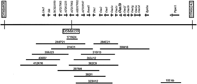
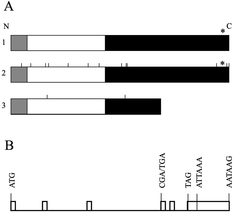
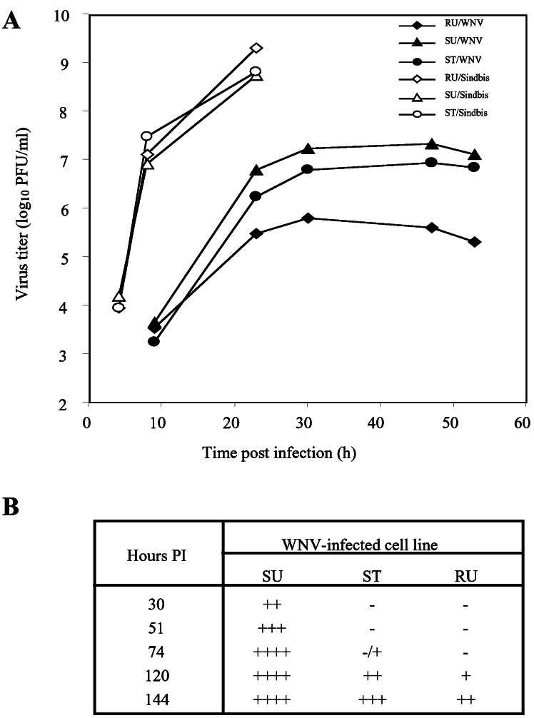
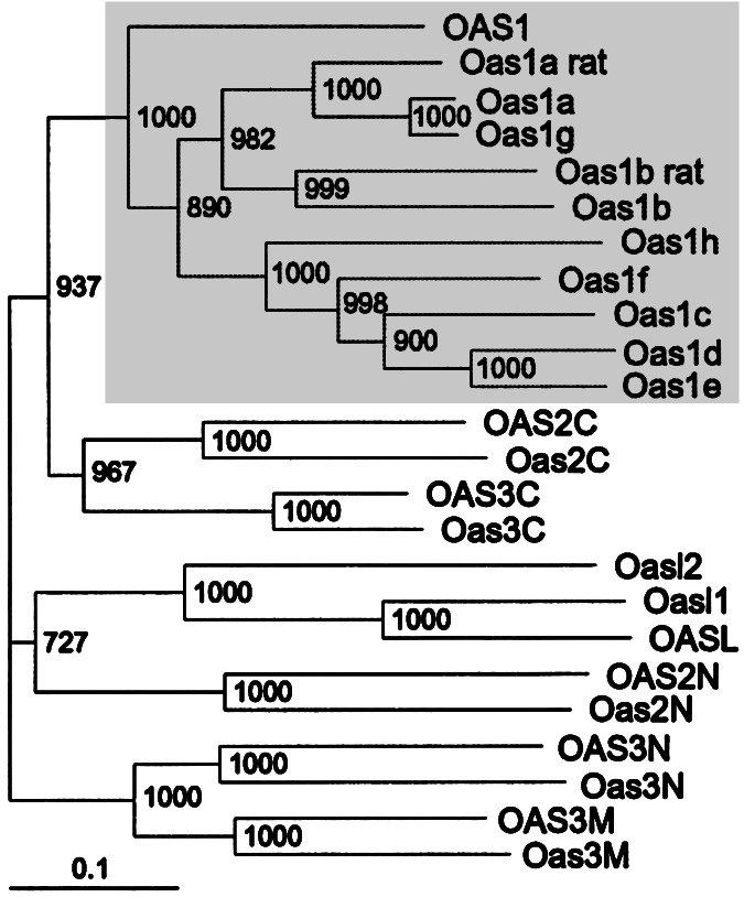
Inbred mouse strains exhibit significant differences in their susceptibility to viruses in the genus Flavivirus, which includes human pathogens such as yellow fever, Dengue, and West Nile virus. A single gene, designated Flv, confers this differential susceptibility and was mapped previously to a region of mouse chromosome 5. A positional cloning strategy was used to identify 22 genes from the Flv gene interval including 10 members of the 2'-5'-oligoadenylate synthetase gene family. One 2'-5'-oligoadenylate synthetase gene, Oas1b, was identified as Flv by correlation between genotype and phenotype in nine mouse strains. Susceptible mouse strains produce a protein lacking 30% of the C-terminal sequence as compared with the resistant counterpart because of the presence of a premature stop codon. The Oas1b gene differs from all the other murine Oas genes by a unique four-amino acid deletion in the P-loop located within the conserved RNA binding domain. Expression of the resistant allele of Oas1b in susceptible embryo fibroblasts resulted in partial inhibition of the replication of a flavivirus but not of an alpha togavirus.


Figures

Figure 1
Figure 1
Physical and transcript maps of the Flv gene interval. Genes are represented by their accepted abbreviations or GenBank accession numbers of their transcripts. The arrows represent the direction of gene transcription. The centromere is oriented toward the left of the figure. The Oas1b gene is indicated in bold. The flanking microsatellite markers are shown inside vertical rectangles, and the D5Mit159 marker is shown inside a horizontal rectangle. The horizontal bars beneath the genes represent the BAC clones listed by their library name.
Figure 2
Figure 2
Structures of the Oas1b gene and protein. (A) Domain architecture of Oas1b proteins. The N-terminal domain (≈30 amino acids, shown in gray) and the C-terminal domain (shown in black) are specific to the Oas protein family (generated with the ProDom tool). The nucleotidyltransferase domain (Pfam 01909) is shown in white. The CFK tetramerization motif is indicated by an asterisk. 1, products of the Flvr and Flvr-like alleles; 2, product of the Flvmr allele; 3, product of the Flvs allele. The positions of amino acid substitutions between the Flvv and Flvmr or the Flvs proteins are shown as vertical bars. (B) Exon-intron structure of the mouse Oas1b gene. Exons are shown as open boxes. The positions of the start (ATG) and stop (TAG) codons, the substitution (CGA/TGA) that results in a premature stop codon, and the two potential polyadenylation sites are indicated by vertical lines.
Figure 3
Figure 3
Constitutive expression of mouse Oas1b in different mouse tissues. A labeled cDNA derived from the 3′ NCR of Oas1bwas used to probe a BALB/c Northern blot (Stratagene) containing poly(A)+ RNA (2 μg per lane) extracted from: 1, heart; 2, kidney; 3, liver; 4, lung; 5, skeletal muscle; or 6, spleen.
Figure 4
Figure 4
Effect of the low level expression of the resistant Oas1b protein in C3H/He cells on the growth of a flavivirus, WNV, and an alpha togavirus, Sindbis. (A) Virus growth curves. Cells were infected with either WNV or Sindbis virus at a multiplicity of infection of 0.5. Samples of culture fluid were taken at the indicated times and titered by plaque assay on BHK cells. RU, untransfected resistant C3H.PRI-Flvr cells; SU, untransfected susceptible C3H/He cells; ST, susceptible C3H/He cells stably transfected with Oas1b cDNA from resistant C3H.PRI-Flvr cells. (B) Time course of the development of CPE after infection of SU, RU, and ST cells with WNV. −, no obvious CPE; +, rounding or detachment of ≈25% of the cells in the monolayer; ++, rounding or detachment of ≈50% of the cells in the monolayer; +++, rounding or detachment of ≈75% of the cells in the monolayer; ++++, complete destruction of the monolayer. PI, postinfection.
Figure 5
Figure 5
Unrooted neighbor-joining distance-based phylogenic tree of murine, rat, and human Oas protein sequences. Human genes are designated by capital letters, but only the first letter is capitalized for the mouse genes. The sequences of the Oas2 and OAS2 proteins were divided into N- and C-terminal domains according to ref. . The sequences of Oas3 and OAS3 proteins were divided into N-terminal (N), middle (M), and C-terminal (C) domains. The indicated bootstrap values were obtained with 1,000 pseudoreplicates. The Oas1 cluster is shown on a gray background. The bar indicates the number of substitutions per site.

Comment in

Similar articles

Cited by

References

    1. Webster L T. J Exp Med. 1923;37:231–244. - PMC - PubMed
    1. Sabin A B. Ann NY Acad Sci. 1952;54:936–944. - PubMed
    1. Green M C. In: Genetic Variants and Strains of the Laboratory Mice. Green MC, editor. Stuttgart: Gustav Fischer; 1981. p. 84.
    1. Shellam G R, Sangster M Y, Urosevic N. Rev Sci Tech. 1998;17:231–248. - PubMed
    1. Goodman G T, Koprowski H. Proc Natl Acad Sci USA. 1962;48:160–165. - PMC - PubMed

Publication types

Substances