. 2002 Jul;184(14):3917-22.
doi: 10.1128/JB.184.14.3917-3922.2002.

Group I self-splicing intron in the recA gene of Bacillus anthracis

Affiliations

Group I self-splicing intron in the recA gene of Bacillus anthracis

Minsu Ko et al. J Bacteriol. 2002 Jul.

Abstract

Self-splicing introns are rarely found in bacteria and bacteriophages. They are classified into group I and II according to their structural features and splicing mechanisms. While the group I introns are occasionally found in protein-coding regions of phage genomes and in several tRNA genes of cyanobacteria and proteobacteria, they had not been found in protein-coding regions of bacterial genomes. Here we report a group I intron in the recA gene of Bacillus anthracis which was initially found by DNA sequencing as an intervening sequence (IVS). By using reverse transcriptase PCR, the IVS was shown to be removable from the recA precursor mRNA for RecA that was being translated in E. coli. The splicing was visualized in vitro with labeled free GTP, indicating that it is a group I intron, which is also implied by its predicted secondary structure. The RecA protein of B. anthracis expressed in E. coli was functional in its ability to complement a recA defect. When recA-negative E. coli cells were irradiated with UV, the Bacillus RecA reduced the UV susceptibility of the recA mutant, regardless of the presence of intron.


Figures

FIG. 1.
FIG. 1.
Schematic diagram of B. anthracis recA gene. The gene includes a 327-bp IVS, located after residue 203. The thick arrows indicate locations of oligonucleotides used. The predicted transcriptional start site and the termination loop are also indicated.
FIG. 2.
FIG. 2.
In vivo and in vitro splicing of recA transcript. (A) In vivo splicing of recA transcript. Lane 1 shows a PCR product obtained from B. anthracis chromosomal DNA with the RECA-4 and RECA-5 primers. Results of RT-PCR from total RNA of B. anthracis using RECA-4 and RECA-5 are also shown, in the absence (lane 2) or presence (lane 3) of reverse transcriptase. Lane M, DNA size markers. (B) Labeling of intron RNA with [α-32P]GTP. Total RNAs were extracted from strains with various plasmids as described in Materials and Methods and incubated with [α-32P]GTP at 37°C in a splicing buffer. Lane 1, BL21/pBluescript SK(+) incubated for 60 min; lane 2 to 5, BL21/pT7recA [int+] incubated for 0 (lane 2), 10 (lane 3), 30 (lane 4), and 60 (lane 5) min; lane 6, BL21/pT7recA [int] incubated for 60 min. After the reaction, samples were analyzed by 5% acrylamide-8 M urea gel electrophoresis and exposed to an X-ray film. The size of detected band was approximately the same as that of the intron (327 bp). A 32P-labeled DNA ladder was used as a marker (M).
FIG. 3.
FIG. 3.
Predicted secondary structure of the recA intron. The arrows indicate the boundaries between exons (lowercase) and intron (uppercase). The conserved base-paired regions (P1 to P9), extra pairings (P1a, P1b, P3.1, P3.2, P6a, and P7.2), and conserved sequence elements (R and S) are shown. The boxed residues, UGG near P1 and CCA near P9, can make a pair (P10) to promote an alignment between the 3′ and 5′ splice sites required for the ligation of exons. The G site (black box) is responsible for the binding of the guanosine cofactor. The shaded boxes represent residues identical with the bnrdE-I1 intron from the Bacillus strain BSG40 (18).
FIG. 4.
FIG. 4.
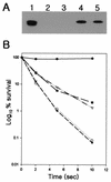
Complementation of recA deletion in E. coli by expression of B. anthracis recA. (A) Bacillus RecA expressed in E. coli. Total proteins were analyzed by immunoblotting with an anti-RecAEC antiserum after sodium dodecyl sulfate-polyacrylamide (12%) gel electrophoresis and transfer to a nitrocellulose paper. Lane 1, MG1655; lane 2, MG1655ΔrecA; lane 3, MG1655ΔrecA/pUC19; lane 4, MG1655ΔrecA /pUCrecA [int+]; lane 5, MG1655ΔrecA/pUCrecA [int]. The size of the B. anthracis RecA protein was about 37 kDa, which is similar to that of E. coli RecA. (B) Complementation of E. coli recA defect by cloned B. anthracis recA. Strains used were MG1655 (•), MG1655ΔrecA (○), MG1655ΔrecA/pUC19 (▾), MG1655ΔrecA/pUCrecA [int+] (▿), and MGΔrecA/pUCrecA [int] (▪). The numbers of colonies were counted after UV irradiation and plating of the samples onto LB agar plates. The plates were incubated for 20 h at 37°C.

Similar articles

Cited by

References

    1. Bechhofer, D. H., K. K. Hue, and D. A. Shub. 1994. An intron in the thymidylate synthase gene of Bacillus bacteriophage B22: evidence for independent evolution of a gene, its group I intron, and the intron open reading frame. Proc. Natl. Acad. Sci. USA 91:11669-11673. - PMC - PubMed
    1. Cech, T. R. 1990. Self-splicing of group I introns. Annu. Rev. Biochem. 59:543-568. - PubMed
    1. Chu, F. K., G. F. Maley, F. Maley, and M. Belfort. 1984. Intervening sequence in the thymidylate synthase gene of bacteriophage T4. Proc. Natl. Acad. Sci. USA 81:3049-3053. - PMC - PubMed
    1. Davies, R. W., R. B. Waring, J. A. Ray, T. A. Brown, and C. Scazzocchio. 1982. Making ends meet: a model for RNA splicing in fungal mitochondria. Nature 300:719-724. - PubMed
    1. Davis, E. O., P. J. Jenner, P. C. Brooks, M. J. Colston, and S. G. Sedgwick. 1992. Protein splicing in the maturation of M. tuberculosis RecA protein: a mechanism for tolerating a novel class of intervening sequence. Cell 71:201-210. - PubMed

Publication types

Substances

LinkOut - more resources