. 2002 Jun 24;157(7):1113-23.
doi: 10.1083/jcb.200112074. Epub 2002 Jun 24.

Cell cycle-dependent localization of macroH2A in chromatin of the inactive X chromosome

Affiliations

Cell cycle-dependent localization of macroH2A in chromatin of the inactive X chromosome

Brian P Chadwick et al. J Cell Biol. .

Abstract

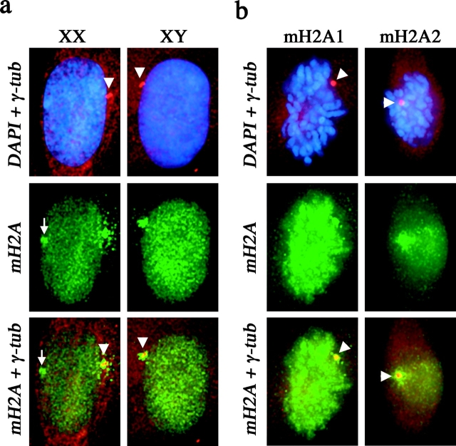
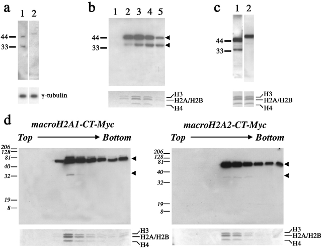
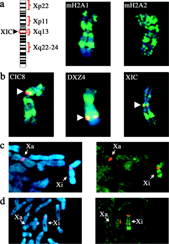
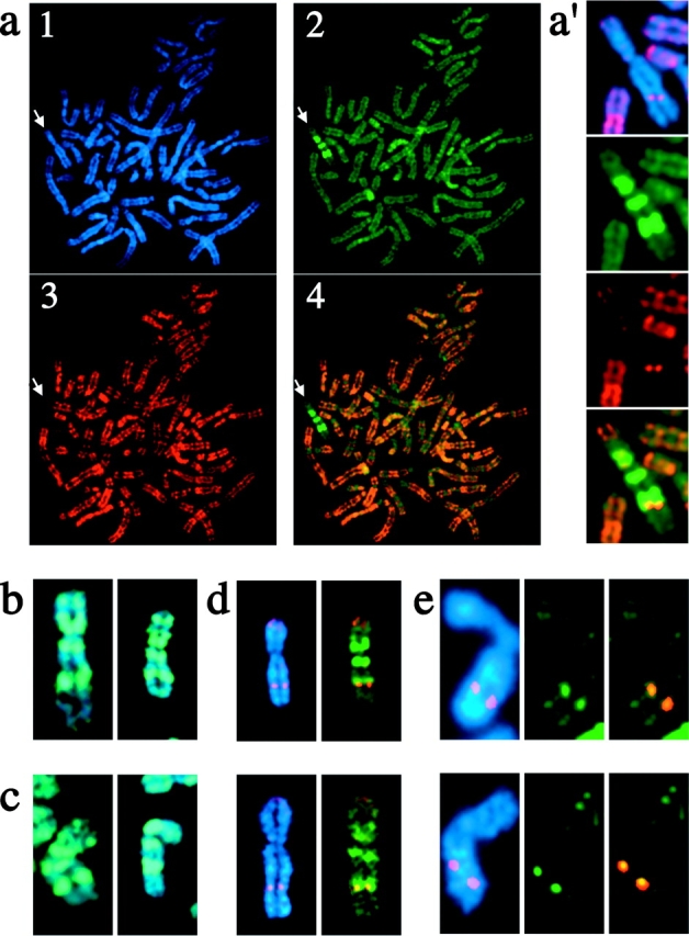
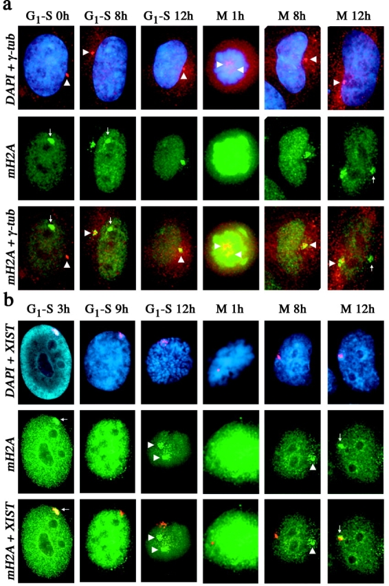
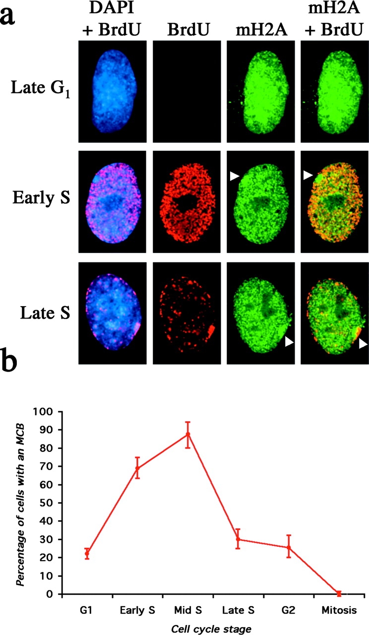
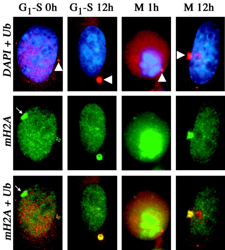
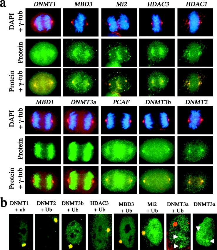
One of several features acquired by chromatin of the inactive X chromosome (Xi) is enrichment for the core histone H2A variant macroH2A within a distinct nuclear structure referred to as a macrochromatin body (MCB). In addition to localizing to the MCB, macroH2A accumulates at a perinuclear structure centered at the centrosome. To better understand the association of macroH2A1 with the centrosome and the formation of an MCB, we investigated the distribution of macroH2A1 throughout the somatic cell cycle. Unlike Xi-specific RNA, which associates with the Xi throughout interphase, the appearance of an MCB is predominantly a feature of S phase. Although the MCB dissipates during late S phase and G2 before reforming in late G1, macroH2A1 remains associated during mitosis with specific regions of the Xi, including at the X inactivation center. This association yields a distinct macroH2A banding pattern that overlaps with the site of histone H3 lysine-4 methylation centered at the DXZ4 locus in Xq24. The centrosomal pool of macroH2A1 accumulates in the presence of an inhibitor of the 20S proteasome. Therefore, targeting of macroH2A1 to the centrosome is likely part of a degradation pathway, a mechanism common to a variety of other chromatin proteins.


Figures

Figure 1.
Figure 1.
Colocalization of macroH2A with centrosomes in somatic cells. (a) Colocalization of macroH2A1 (green, FITC) with γ-tubulin (red, TRITC) in 46,XX (T-3352) and 46,XY (hTERT-BJ1) cells. Nuclei are stained with DAPI (blue). The white arrowhead indicates the position of the centrosome, determined by γ-tubulin staining. The merged macroH2A1 and γ-tubulin signals indicate overlapping signals in yellow. The white arrow indicates the location of the MCB (green) in the 46,XX nucleus. (b) Colocalization of macroH2A1 and macroH2A2 with γ-tubulin in 46,XX (hTERT-RPE1) cells by indirect immunofluorescence. Cells grown directly on microscope slides were stained with either anti-macroH2A1 or anti-macroH2A2 (aa 124–180). Metaphase chromosomes are stained with DAPI. The white arrowhead indicates the position of the centrosome, determined by γ-tubulin staining. The merged macroH2A and γ-tubulin signals indicate overlapping signals in yellow.
Figure 2.
Figure 2.
Detection of macroH2A1 and macroH2A2 in fractions from nucleosome and centrosome preparations. (a) Immunoblot analysis of the same fraction of a centrosome preparation from a 46,XX (hTERT-RPE1) cell line detecting the presence of macroH2A1 (lane 1) or macroH2A2 (aa 124–372; lane 2). The panel below shows the relative level of γ-tubulin by immunoblotting in the same fraction. (b) Immunoblot of five consecutive sucrose gradient fractions (lanes 1–5) of a nucleosome preparation from a 46,XX (hTERT-RPE1) cell line, showing the cofractionation of full-length macroH2A1 (top arrowhead) and a smaller signal (bottom arrowhead). The relative concentration of nucleosomes/histones in each fraction is shown in the Coomassie image below. (c) Immunoblot analysis of the same nucleosome-containing fraction of a nucleosome preparation from a 46,XX (hTERT-RPE1) cell line detecting the presence of macroH2A1 (lane 1) or macroH2A2 (aa 124–372; lane 2). The panel below shows an image of the Coomassie stain of the same fraction indicating the positions of the histones. (d) Immunoblot analysis using anti-Myc mAb detecting the presence of macroH2A1-CT–Myc and macroH2A2-CT–Myc in sucrose gradient fractions of nucleosome preparations from macroH2A1-CT–Myc- and macroH2A2-CT–Myc-expressing HEK-293 cell lines. The arrowheads to the right of each image indicate the presence of the upper full-length macroH2A1/2–Myc and a smaller macroH2A1/2–Myc-derived form. The relative concentration of nucleosomes/histones in each fraction is shown in the Coomassie image below.
Figure 3.
Figure 3.
MacroH2A displays distinct banding patterns on the metaphase Xi chromosome. (a) 46,XX (hTERT-RPE1) metaphase Xi chromosome stained with macroH2A1- or macroH2A2- (aa 124–180) specific pAb by indirect immunofluorescence (green, FITC), merged with DAPI staining (blue). The ideogram represents the metaphase Xi chromosome indicating the position of the X inactivation center (XIC) and the positions of the four main bands at Xp22, Xp11, Xq13, and Xq22-24. (b) Indirect immunofluorescence of macroH2A1 banding pattern on the Xi chromosome in 46,XX (hTERT-RPE1) cells merged with FISH signals (red, TRITC) for ordered X chromosome probes, and merged with DAPI image. The white arrowheads indicate the location of the specific FISH probes. Overlapping signals are in yellow. (c) Partial metaphase spread from a 46,XX (hTERT-HME1) cell line stained with macroH2A1 by indirect immunofluorescence, merged with X-α satellite FISH signals. The Xa and Xi are indicated. (d) Partial metaphase spread from a female mouse cell line (B144) stained with macroH2A1 by indirect immunofluorescence, merged with X chromosome–specific DXwas70 FISH signals. The Xa and Xi are indicated.
Figure 4.
Figure 4.
Overlap of the Xq22-24 macroH2A1 band with a band of histone H3 lysine-4 methylation centered at DXZ4. (a) Female metaphase chromosomes from a macroH2A1-CT–Myc-transfected 46,XX (hTERT-RPE1) cell line counterstained with anti-DimH3K4. Chromosomes are stained with DAPI (blue; panel 1), and the distribution of macroH2A1 (green, FITC; panel 2) and DimH3K4 (red, TRITC; panel 3) are shown. The merged distribution of macroH2A1 and DimH3K4 are shown in panel 4 with overlapping signals in orange. White arrow points to the position of the Xi. (a′) Enlarged image of the Xi indicated, showing the macroH2A1 and DimH3K4 banding patterns on the Xi. The DAPI image is merged with the DimH3K4 signal in the top panel to indicate the relative position of the DimH3K4 signal on Xq. (b) 46,XX (hTERT-RPE1) Xi chromosomes showing the distribution of macroH2A1-CT–Myc, merged with the DAPI image. The distribution is indistinguishable from that seen for the endogenous macroH2A1 protein (Fig. 2). (c) 46,XX (hTERT-RPE1) Xi chromosomes showing the distribution of macroH2A1-NT–Xpr, merged with the DAPI image. (d) 46,XX (hTERT-RPE1) Xi chromosomes showing the distribution of DimH3K4 merged with either the DAPI image or the distribution of macroH2A1-CT–Myc, with overlapping signals shown in yellow. (e) 46,XX (hTERT-RPE1) Xi chromosomes showing the DXZ4 locus, merged with the DAPI image or the DimH3K4 signal. Overlapping DXZ4 and DimH3K4 signals are shown in yellow.
Figure 5.
Figure 5.
Distribution of macroH2A1 in relation to the centrosome and XIST RNA through the somatic cell cycle. (a) Distribution of macroH2A1 (green, FITC) and γ-tubulin (red, TRITC) in 46,XX (hTERT-RPE1) cells at different stages of the cell cycle as detected by indirect immunofluorescence. The nucleus is stained by DAPI (blue). Overlapping signals are shown in yellow. The positions of the centrosome (white arrowhead) and MCB (white arrow) are indicated. The stage of the cell cycle from which cells were released, G1–S or mitosis (M), are indicated along with the number of hours (h) after release. (b) Distribution of macroH2A1 and XIST RNA in 46,XX (hTERT-RPE1) cells at different stages of the cell cycle as detected by indirect immunofluorescence and RNA-FISH. The nucleus is stained by DAPI. Overlapping signals are shown in yellow. The position of the centrosome (white arrowhead) and MCB (white arrow) are indicated. The stage of the cell cycle from which cells were released, G1–S or mitosis (M), are indicated along with the number of hours (h) after release.
Figure 6.
Figure 6.
Distribution of macroH2A1 in synchronized 46,XX (hTERT-RPE1) cells in different stages of the somatic cell cycle. (a) The late G1 cell was released from mitosis for 14 h and pulsed with BrdU for 1 h before detection of macroH2A1 (green, FITC) and BrdU (red, TRITC). The nucleus is stained with DAPI (Blue). The cell has not yet entered S phase, as determined by the lack of an anti-BrdU signal. The early S phase cell was released from mitosis for 16 h and pulsed with BrdU for 1 h before detection of macroH2A and BrdU. The nucleus is stained with DAPI and merged with the anti-BrdU signal. The position of the MCB is indicated by the white arrowhead and, as indicated by the merged macroH2A1 and BrdU signals (yellow), has not yet undergone DNA replication. The late S phase cell was released from G1–S for 9 h and pulsed with BrdU for 1 h before detection of macroH2A1 and BrdU. The nucleus is stained with DAPI and merged with the anti-BrdU signal. The position of the MCB is indicated by the white arrowhead and, as indicated by the merged macroH2A1 and BrdU signals (yellow), is currently undergoing DNA replication. (b) Graph showing the frequency of MCB observation at different stages of the somatic cell cycle. Synchronized 46,XX cells (hTERT-RPE1) released for different time periods from the G1–S boundary or mitosis and pulsed for 1 h with BrdU. Cells were scored for the presence of an MCB at different cell cycle stages.
Figure 7.
Figure 7.
Colocalization of macroH2A1 with ubiquitin and accumulation at the centrosomal proteasome over time with inhibition of the 20S proteasome. 46,XX (hTERT-RPE1) cells were synchronized at G1–S or mitosis (M) and directly processed (0 h/1 h) or released for 12 h (12 h) in the presence of 10 μM lactacystin. The distribution of macroH2A1 (green, FITC) (mH2A) and ubiquitin (red, TRITC) (Ub) are shown by indirect immunofluorescence. The nucleus is stained with DAPI (blue). The white arrowheads indicate the position of the centrosome proteasome marked by ubiquitin, and the small white arrows indicate the position of MCBs. Overlapping macroH2A1 and ubiquitin (yellow) show colocalization and accumulation 12 h after G1–S and mitosis in the presence of the proteasome inhibitor lactacystin.
Figure 8.
Figure 8.
Colocalization of a number of heterochromatin proteins with centrosomes at mitosis, and accumulation at the centrosome proteasome in the presence of lactacystin. (a) Indirect immunofluorescence of heterochromatin proteins (green, FITC) with γ-tubulin (red, TRITC) in 46,XX (hTERT-RPE1) at mitosis. Chromosomes are stained with DAPI (blue). Overlapping heterochromatin protein signals and γ-tubulin signals in merged images are indicated in yellow. The different heterochromatin proteins are indicated to the left of each group of panels. (b) Extensive incubation of 46,XX (hTERT-RPE1) cells with lactacystin results in the accumulation of some, but not all, heterochromatin proteins at the centrosome proteasome. Cells were treated with 10 μM lactacystin for 14 h before processing. Ubiquitin and proteins indicated were detected by indirect immunofluorescence. Clear overlapping signals with ubiquitin can be seen (yellow). White arrowheads indicate the accumulation of Dnmt3a with two nuclear territories and not with the centrosome proteasome. Gene names are defined in the Materials and methods.

References

    1. Avner, P., and E. Heard. 2001. X-chromosome inactivation: counting, choice and initiation. Nat. Rev. Genet. 2:59–67. - PubMed
    1. Barthelmes, H.U., P. Grue, S. Feineis, T. Straub, and F. Boege. 2000. Active DNA topoisomerase IIα is a component of the salt-stable centrosome core. J. Biol. Chem. 275:38823–38830. - PubMed
    1. Beletskii, A., Y.-K. Hong, J. Pehrson, M. Egholm, and W.M. Strauss. 2001. PNA interference mapping demonstrates functional domains in the noncoding RNA Xist. Proc. Natl. Acad. Sci. USA. 98:9215–9220. - PMC - PubMed
    1. Belyaev, N., A.M. Keohane, and B.M. Turner. 1996. Differential underacetylation of histones H2A, H3 and H4 on the inactive X chromosome in human female cells. Hum. Genet. 97:573–578. - PubMed
    1. Bochtler, M., L. Ditzel, M. Groll, C. Hartmann, and R. Huber. 1999. The proteasome. Annu. Rev. Biophys. Biomol. Struct. 28:295–317. - PubMed

Publication types