. 2002 Jun 24;157(7):1139-49.
doi: 10.1083/jcb.200203035. Epub 2002 Jun 24.

The roles of Fzy/Cdc20 and Fzr/Cdh1 in regulating the destruction of cyclin B in space and time

Affiliations

The roles of Fzy/Cdc20 and Fzr/Cdh1 in regulating the destruction of cyclin B in space and time

Jordan W Raff et al. J Cell Biol. .

Abstract

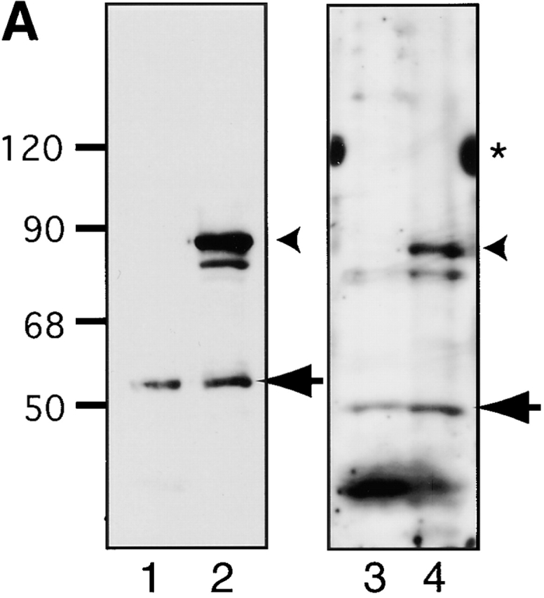
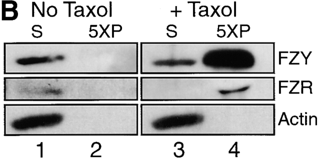
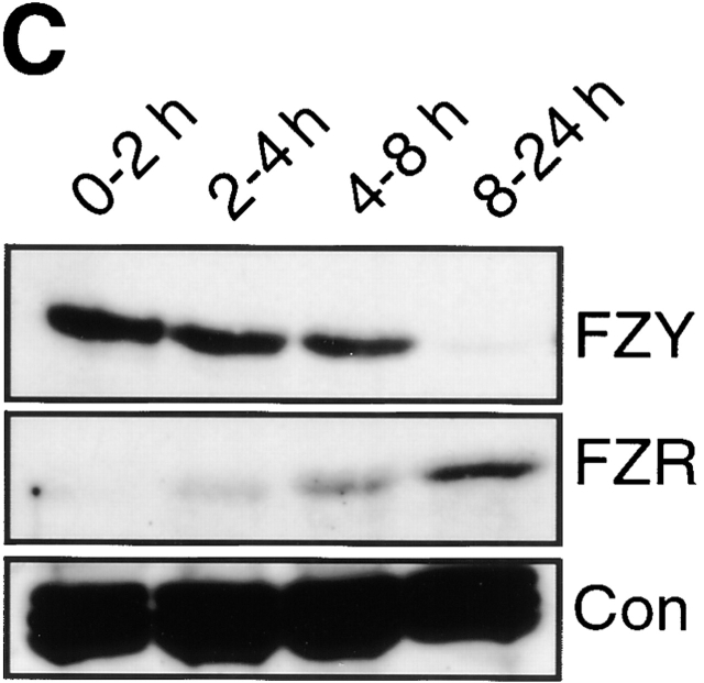
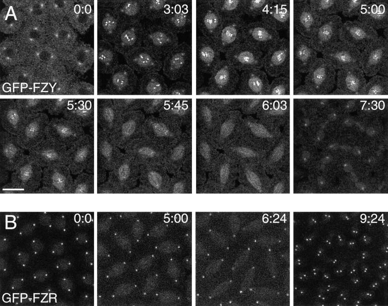
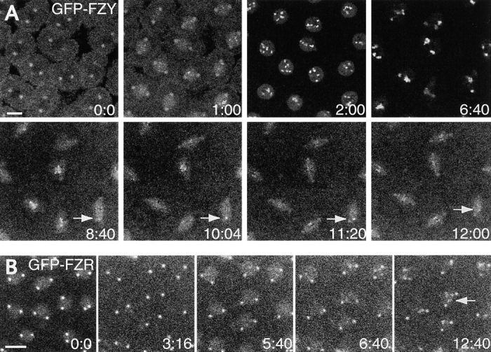
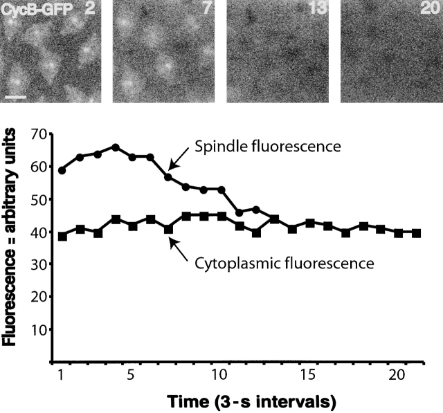
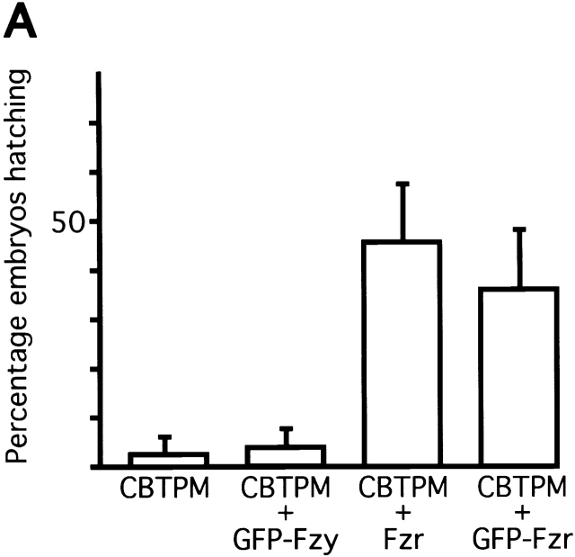
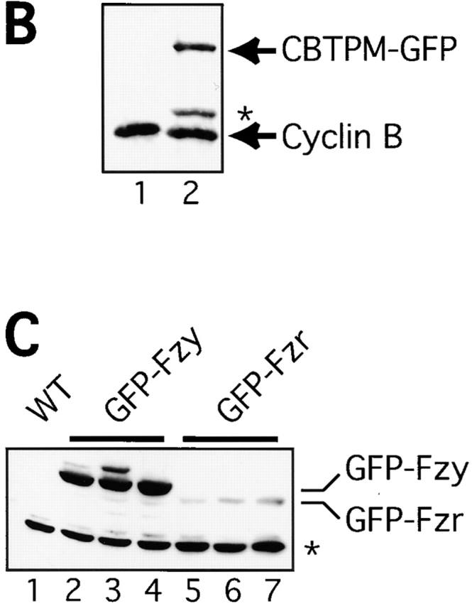
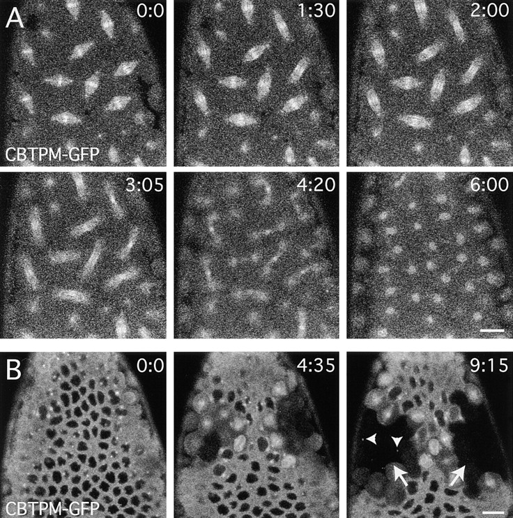
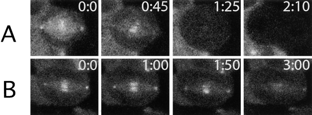
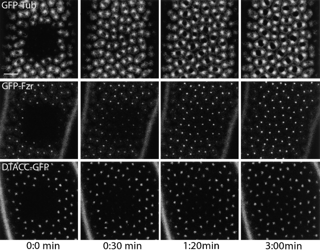
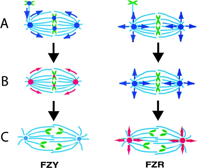
In Drosophila cells cyclin B is normally degraded in two phases: (a) destruction of the spindle-associated cyclin B initiates at centrosomes and spreads to the spindle equator; and (b) any remaining cytoplasmic cyclin B is degraded slightly later in mitosis. We show that the APC/C regulators Fizzy (Fzy)/Cdc20 and Fzy-related (Fzr)/Cdh1 bind to microtubules in vitro and associate with spindles in vivo. Fzy/Cdc20 is concentrated at kinetochores and centrosomes early in mitosis, whereas Fzr/Cdh1 is concentrated at centrosomes throughout the cell cycle. In syncytial embryos, only Fzy/Cdc20 is present, and only the spindle-associated cyclin B is degraded at the end of mitosis. A destruction box-mutated form of cyclin B (cyclin B triple-point mutant [CBTPM]-GFP) that cannot be targeted for destruction by Fzy/Cdc20, is no longer degraded on spindles in syncytial embryos. However, CBTPM-GFP can be targeted for destruction by Fzr/Cdh1. In cellularized embryos, which normally express Fzr/Cdh1, CBTPM-GFP is degraded throughout the cell but with slowed kinetics. These findings suggest that Fzy/Cdc20 is responsible for catalyzing the first phase of cyclin B destruction that occurs on the mitotic spindle, whereas Fzr/Cdh1 is responsible for catalyzing the second phase of cyclin B destruction that occurs throughout the cell. These observations have important implications for the mechanisms of the spindle checkpoint.


Figures

Figure 1.
Figure 1.
The behavior of Fzy/Cdc20 and Fzr/Cdh1 in Western blotting experiments. (A) WT embryos (lanes 1 and 3) or embryos expressing either GFP-Fzy (lane 2) or GFP-Fzr (lane 4) were probed with affinity-purified anti-Fzy (lanes 1 and 2) or anti-Fzr (lanes 3 and 4) antibodies. Arrows highlight the position of Fzy or Fzr; arrowheads highlight the position of GFP-Fzy or GFP-Fzr. The anti-Fzr antibodies also recognize several other proteins in the embryo extract, as well as the 120-kD marker protein (asterisk). The position of marker proteins is indicated at the left of the figure. (B) A microtubule spindown experiment probed with anti-Fzy (top), anti-Fzr (middle), or anti-actin (bottom) antibodies. In control experiments, where no taxol is added, all three proteins remain in the extract supernatant (S, lane 1) and are not present in the pellet (5 × P, lane 2; the 5× indicates that 5× more of the pellet was loaded on the gel relative to the supernatant). If taxol is added to the embryo extracts, a significant fraction (∼50–70%) of Fzy/Cdc20 and Fzr/Cdh1 copellet with the microtubules, whereas actin does not (lane 4). (C) The developmental expression of Fzy (top) and Fzr (bottom) proteins. Equal numbers of 0–2-, 2–4-, 4–8-, or 8–24-h-old embryos were loaded in each lane. The bottom panel shows the levels of a nonspecific band recognized by the Fzr antibodies, shown here as a loading control.
Figure 1.
Figure 1.
The behavior of Fzy/Cdc20 and Fzr/Cdh1 in Western blotting experiments. (A) WT embryos (lanes 1 and 3) or embryos expressing either GFP-Fzy (lane 2) or GFP-Fzr (lane 4) were probed with affinity-purified anti-Fzy (lanes 1 and 2) or anti-Fzr (lanes 3 and 4) antibodies. Arrows highlight the position of Fzy or Fzr; arrowheads highlight the position of GFP-Fzy or GFP-Fzr. The anti-Fzr antibodies also recognize several other proteins in the embryo extract, as well as the 120-kD marker protein (asterisk). The position of marker proteins is indicated at the left of the figure. (B) A microtubule spindown experiment probed with anti-Fzy (top), anti-Fzr (middle), or anti-actin (bottom) antibodies. In control experiments, where no taxol is added, all three proteins remain in the extract supernatant (S, lane 1) and are not present in the pellet (5 × P, lane 2; the 5× indicates that 5× more of the pellet was loaded on the gel relative to the supernatant). If taxol is added to the embryo extracts, a significant fraction (∼50–70%) of Fzy/Cdc20 and Fzr/Cdh1 copellet with the microtubules, whereas actin does not (lane 4). (C) The developmental expression of Fzy (top) and Fzr (bottom) proteins. Equal numbers of 0–2-, 2–4-, 4–8-, or 8–24-h-old embryos were loaded in each lane. The bottom panel shows the levels of a nonspecific band recognized by the Fzr antibodies, shown here as a loading control.
Figure 2.
Figure 2.
The behavior of GFP-Fzy and GFP-Fzr in living syncytial embryos. The GFP-Fzy (A)– and GFP-Fzr (B)–expressing embryos shown here were followed from interphase to telophase (see text for details). Time in minutes is shown at the top right of each panel. Bar, 20 μm.
Figure 3.
Figure 3.
The behavior of GFP-Fzy and GFP-Fzr in living embryos injected with colcemid. Embryos were injected in interphase and they then arrested in a mitotic state (see text for details). The GFP-Fzy expressing embryo was then subjected to a single pulse of UV light at 7:00 min to inactivate the colcemid. The arrow in A highlights the position of a kinetochore that is delayed in lining up on the metaphase plate of the reforming spindle, and retains high levels of GFP-Fzy until it does so. The arrow in B highlights the GFP-Fzr dots that appear to associate with the chromosomes as the embryos arrest in mitosis. Time in minutes is shown in the bottom right of each panel. Bars, 20 μm.
Figure 4.
Figure 4.
The destruction of cyclin B–GFP in living syncytial embryos. Selected images of a syncytial embryo exiting mitosis are shown here. The graph shows the quantitation (Materials and methods) of fluorescence intensity on the spindle (•) and in the cytoplasm (▪) measured at 3-s intervals as the embryo exits mitosis. The number in the top right-hand corner of each image corresponds to the number on the time line of the graph. Bar, 20 μm.
Figure 5.
Figure 5.
The expression of Fzr/Cdh1 or GFP-Fzr partially rescues the lethality associated with expressing CBTPM–GFP in early embryos. (A) A graph showing the percentage of embryos that hatch when CBTPM–GFP alone is expressed in embryos, or when CBTPM–GFP is coexpressed with GFP-Fzy, GFP-Fzr, or Fzr alone (as indicated below each bar). Error bars represent the standard deviation. (B) A Western blot showing the relative expression level of CBTPM–GFP in syncytial embryos compared with the endogenous cyclin B. Equal numbers of WT (lane 1) or CBTPM–GFP-expressing (lane 2) embryos were loaded in each lane. Arrows show the position of the endogenous cyclin B and of the CBTPM–GFP. The asterisk shows the position of a prominent breakdown product of CBTPM–GFP. (C) A Western blot showing the relative levels of expression of GFP-Fzy and GFP-Fzr. Equal numbers of syncytial embryos from three different lines expressing GFP-Fzy (lanes 2–4), GFP-Fzr (lanes 5–7), or a WT control (lane 1) were probed with anti GFP antibodies. The asterisk marks a crossreacting band that is recognized by the GFP antibodies in embryo extracts, shown here as a loading control. In all lines tested, the GFP-Fzy protein is expressed at ∼10-fold higher levels than GFP-Fzr.
Figure 5.
Figure 5.
The expression of Fzr/Cdh1 or GFP-Fzr partially rescues the lethality associated with expressing CBTPM–GFP in early embryos. (A) A graph showing the percentage of embryos that hatch when CBTPM–GFP alone is expressed in embryos, or when CBTPM–GFP is coexpressed with GFP-Fzy, GFP-Fzr, or Fzr alone (as indicated below each bar). Error bars represent the standard deviation. (B) A Western blot showing the relative expression level of CBTPM–GFP in syncytial embryos compared with the endogenous cyclin B. Equal numbers of WT (lane 1) or CBTPM–GFP-expressing (lane 2) embryos were loaded in each lane. Arrows show the position of the endogenous cyclin B and of the CBTPM–GFP. The asterisk shows the position of a prominent breakdown product of CBTPM–GFP. (C) A Western blot showing the relative levels of expression of GFP-Fzy and GFP-Fzr. Equal numbers of syncytial embryos from three different lines expressing GFP-Fzy (lanes 2–4), GFP-Fzr (lanes 5–7), or a WT control (lane 1) were probed with anti GFP antibodies. The asterisk marks a crossreacting band that is recognized by the GFP antibodies in embryo extracts, shown here as a loading control. In all lines tested, the GFP-Fzy protein is expressed at ∼10-fold higher levels than GFP-Fzr.
Figure 6.
Figure 6.
The behavior of CBTPM–GFP in living syncytial or cellularized embryos. (A) CBTPM–GFP usually arrests syncytial embryos in mitosis (Wakefield et al., 2000). However, in the syncytial embryo shown here, the spindles exit mitosis but CBTPM–GFP remains concentrated on centrosomes and spindles throughout this time (WT cyclin B–GFP is normally degraded on the spindle at this time; Fig. 4). (B) In a cellularized embryo, CBTPM–GFP is degraded at the end of mitosis (the arrows highlight the position of two mitotic domains that have exited mitosis). However, the kinetics of degradation are not normal, and the spindle remnants still contain CBTPM–GFP even after mitosis is finished (arrowheads). Time in minutes is shown in the top right of each panel. Bars, 10 μm.
Figure 7.
Figure 7.
A comparison of the destruction of cyclin B–GFP and CBTPM–GFP in cellularized embryos. Whereas cyclin B–GFP (A) is no longer detectable on spindles as they enter anaphase, CBTPM–GFP (B) can still be detected on anaphase spindles. Quantitation of the fluorescence levels in these cells (Materials and methods) revealed that both fusion proteins were eventually completely degraded in both cells, apart from a small amount of protein that remained on the spindle remnants of the CBTPM–GFP-expressing cell (unpublished data; Fig. 6 B). The number in the top right-hand corner corresponds to the time in minutes.
Figure 8.
Figure 8.
GFP-Fzr is rapidly turned over at centrosomes. Living embryos expressing GFP-tubulin (A), GFP-Fzr (B), or D-TACC-GFP (C) were followed on the confocal microscope. During interphase of nuclear cycle 10–12, a small region of the embryo was bleached with 100% laser power, and the recovery of fluorescence was followed. GFP-tubulin recovers quickly (T1/2 ∼10 s, Materials and methods); GFP-Fzr recovers at an intermediate rate (T1/2 ∼45 s); D-TACC-GFP recovers more slowly (T1/2 ∼2 min). Time, in minutes, after photobleaching is indicated at the bottom of the figure. Bar, 20 μm.
Figure 9.
Figure 9.
A model of how the sequential activation of Fzy/Cdc20 and Fzr/Cdh1 regulates the destruction of cyclin B in space and time. Chromosomes are shown in green and microtubules in light blue. When Fzy/Cdc20 (left) and Fzr/Cdh1 (right) are inactivate they are shown as dark blue, and when activated to degrade cyclin B they are shown as red. The motion of these molecules is depicted with arrows. (A) Early in mitosis, inactive Fzy (Cdc20)/checkpoint protein complexes are formed at unattached kinetochores, transported toward the centrosome, and then spread throughout the spindle. Fzr/Cdh1 constantly turns over at centrosomes, (indicated by the arrows showing the protein leaving the centrosome) but is inactive as cyclin B/cdc2 activity is high. (B) Once all the chromosomes align at the metaphase plate, inhibitory complexes of Fzy (Cdc20)/checkpoint proteins are no longer generated at kinetochores and Fzy/Cdc20 is activated to degrade cyclin B at centrosomes (or perhaps at kinetochores). The activated Fzy/Cdc20 complexes spread along the spindle microtubules degrading cyclin B in a wave that appears to spread from the centrosome to the spindle equator. (C) The destruction of cyclin B lowers cdc2 kinase activity at the centrosome, leading to the activation of the Fzr/Cdh1 at the centrosome. The active Fzr/Cdh1 complexes are not restricted to microtubules, and can degrade cyclin B throughout the cell.

Similar articles

Cited by

References

    1. Abrieu, A., J.A. Kahana, K.W. Wood, and D.W. Cleveland. 2000. CENP-E as an essential component of the mitotic checkpoint in vitro. Cell. 102:817–826. - PubMed
    1. Abrieu, A., L. Magnaghi-Jaulin, J.A. Kahana, M. Peter, A. Castro, S. Vigneron, T. Lorca, D.W. Cleveland, and J.C. Labbe. 2001. Mps1 is a kinetochore-associated kinase essential for the vertebrate mitotic checkpoint. Cell. 106:83–93. - PubMed
    1. Bardin, A.J., R. Visintin, and A. Amon. 2000. A mechanism for coupling exit from mitosis to partitioning of the nucleus. Cell. 102:21–31. - PubMed
    1. Basto, R., R. Gomes, and R.E. Karess. 2000. Rough deal and Zw10 are required for the metaphase checkpoint in Drosophila. Nat. Cell Biol. 2:939–943. - PubMed
    1. Chan, G.K., S.A. Jablonski, D.A. Starr, M.L. Goldberg, and T.J. Yen. 2000. Human Zw10 and ROD are mitotic checkpoint proteins that bind to kinetochores. Nat. Cell Biol. 2:944–947. - PubMed

Publication types

MeSH terms