Comparative Study
. 2002 Jun 24;157(7):1279-90.
doi: 10.1083/jcb.200203073. Epub 2002 Jun 24.

Matrix assembly, regulation, and survival functions of laminin and its receptors in embryonic stem cell differentiation

Affiliations
Comparative Study

Matrix assembly, regulation, and survival functions of laminin and its receptors in embryonic stem cell differentiation

Shaohua Li et al. J Cell Biol. .

Abstract

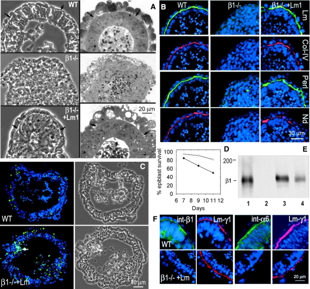
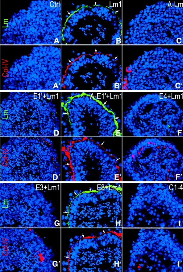
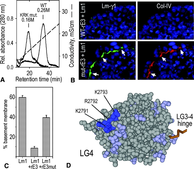
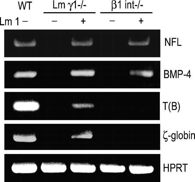
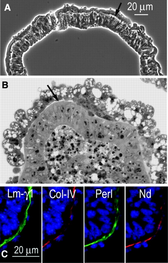
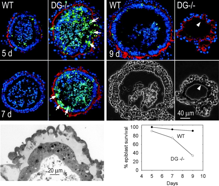
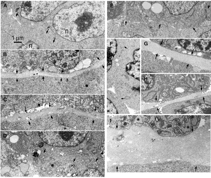
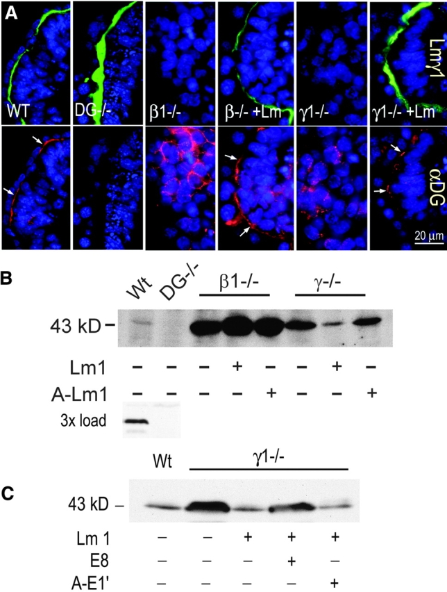
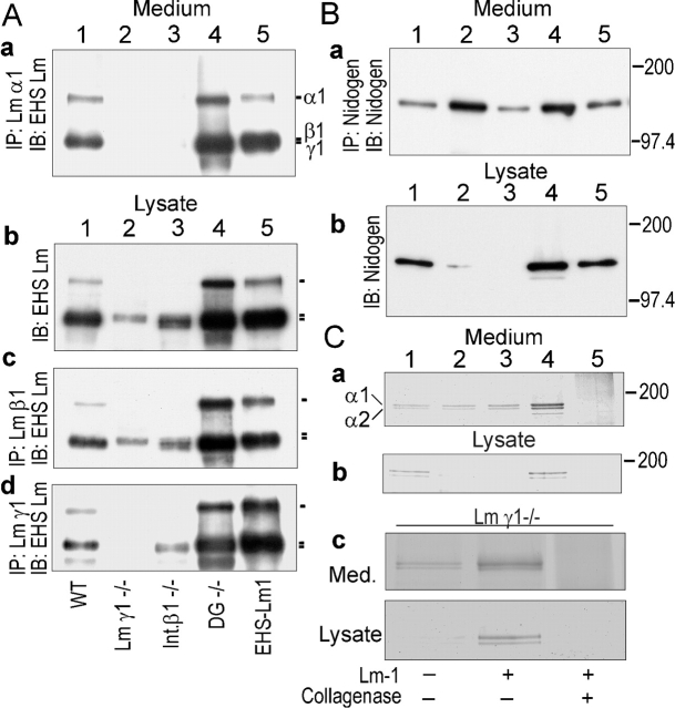
Laminin-1 is essential for early embryonic basement membrane assembly and differentiation. Several steps can be distinguished, i.e., the expression of laminin and companion matrix components, their accumulation on the cell surface and assembly into basement membrane between endoderm and inner cell mass, and the ensuing differentiation of epiblast. In this study, we used differentiating embryoid bodies derived from mouse embryonic stem cells null for gamma1-laminin, beta1-integrin and alpha/beta-dystroglycan to dissect the contributions of laminin domains and interacting receptors to this process. We found that (a) laminin enables beta1-integrin-null embryoid bodies to assemble basement membrane and achieve epiblast with beta1-integrin enabling expression of the laminin alpha1 subunit; (b) basement membrane assembly and differentiation require laminin polymerization in conjunction with cell anchorage, the latter critically dependent upon a heparin-binding locus within LG module-4; (c) dystroglycan is not uniquely required for basement membrane assembly or initial differentiation; (d) dystroglycan and integrin cooperate to sustain survival of the epiblast and regulate laminin expression; and (e) laminin, acting via beta1-integrin through LG1-3 and requiring polymerization, can regulate dystroglycan expression.


Figures

Figure 1.
Figure 1.
Laminin induction of basement membrane and epiblast in β1-integrin–null EBs. Wild-type and β1-integrin–null ES cells were grown in suspension for 7 d, the latter maintained alone or in the presence of laminin-1 (25 μg/ml). (A) Phase micrographs (left) and methylene blue-stained sections (right) of wild-type (top), untreated (middle) and laminin-1–treated (bottom) β1-integrin–null EBs. (B) Immunofluorescence micrographs of consecutive sections of the above EBs stained with DAPI (nuclei, blue) and antibody to laminin-γ1 (Lm), type IV collagen (Col-IV), perlecan (Perl) and nidogen (Nd). (C and D) β1-integrin–null EBs were treated with 25 μg/ml laminin-1 and cultured for 7–11 d. TUNEL (green) and DAPI (blue) costaining revealed developing segmental (arrow) apoptosis (plot was calculated after subtracting EBs with full-thickness segmental apoptosis from the total). (E) Immunoblot detection of the β1-integrin subunit in wild-type (lane 1), β1-integrin–null (lane 2), dystroglycan-null (lane 3), and γ1-laminin–null (lane 4) EBs. (F) Immunofluorescence micrographs showing β1-integrin (first frame), α6-integrin (third frame), and γ1-laminin (second and fourth frames) of wild-type (top) and laminin-treated β1-integrin–null EBs (bottom).
Figure 2.
Figure 2.
Laminin domains contributing to basement membrane assembly. γ1-Laminin–null EBs were cultured for 7 d without laminin as a control (Ctrl), or with either laminin-1 (Lm1, 25 μg/ml), nonpolymerizing laminin-1 (A-Lm1, AEBSF-treated, 25 μg/ml), or fragment C1–4 (25 μg/ml) as shown. In addition, EBs were treated with laminin-1 mixed with an ∼50-fold molar excess of polymer-inhibiting fragment E1', noninhibiting AEBSF-E1' control (A-E1'), polymer-inhibiting fragment E4, α6β1-integrin–binding fragment E8, or dystroglycan/heparin/sulfatide-binding fragment E3 (fragment map shown in Fig. 10).
Figure 3.
Figure 3.
Heparin-binding site in LG4 is required by laminin mediation of basement membrane assembly in laminin γ1-null EBs. (A) Elution of recombinant wild-type and KRK→AAA mutant recombinant LG4–5 from a heparin 5PW column with a NaCl gradient. (B) Immunofluorescence (laminin γ1, type IV collagen) micrographs of γ1-laminin–null EBs treated with either laminin-1 in the presence of a 20-fold molar excess of wild-type or mutated LG4–5. (C) Quantitation of degree of basement membrane formation. (D) Space-filling model of Lm-α1 LG4 KRK sequence (blue) superimposed upon the crystal structure of α2-LG4 determined from coordinates submitted to the Brookhaven protein database.
Figure 4.
Figure 4.
Expression of differentiation markers. β1-integrin–null (β1 int−/−) and γ1-laminin–null (Lm γ1−/−) ES cells untreated or treated with laminin-1 (Lm1, 25 μg/ml) and wild-type ES cells were cultured in suspension for 7 d. Total RNA was isolated and subjected to semi-quantitative RT-PCR analysis for epiblast (low molecular weight NFL) and mesoderm (brachyury, T(B), and ζ-globin) marker expression. HPRT was used as normalizing control.
Figure 5.
Figure 5.
Basement membranes assemble in dystroglycan-null EBs. Dystroglycan-null ES cells, suspended at the first passage from feeder cell layers, were allowed to form EBs for 5 d in the absence of any treatment. (A) Phase micrograph and (B) methylene blue–stained section show epiblast differentiation, cavitation, and thin basement membranelike structures (arrows). (C) EBs, visualized by immunofluorescence, reveal a subendodermal basement membrane pattern costaining with antibodies for γ1-laminin, type IV collagen, perlecan, and nidogen.
Figure 6.
Figure 6.
Development of epiblast apoptosis and basement membrane thickening in dystroglycan-null EBs. Untreated dystroglycan-null EBs and wild-type controls were cultured for 5–9 d. Thick basement membranes developed in dystroglycan-null EBs as seen both in immunofluorescence micrographs (laminin-γ1 epitope in red) and the thick section. An epiblast layer is seen in both forms of EBs. By 9 d, epiblast layer degeneration is observed. TUNEL (green) and DAPI (blue) costaining reveals apoptosis in wild-type and dystroglycan-null cells over time. Epiblast apoptosis was prominent in the dystroglycan-null, but not the wild-type, EBs, and was augmented over time. The EBs with partial and complete degeneration and loss of the epiblast layer were subtracted from the total EBs counted. The plot shows the percentage of remaining EBs with surviving epiblast layers.
Figure 7.
Figure 7.
Ultrastructure of EBs. Wild-type (R1), γ1-laminin–null, β1-integrin–null, and dystroglycan-null EBs, untreated and laminin-treated, were examined by electron microscopy after incubation in suspension culture for 5–7 d. The regions containing junctions of the endodermal layer and ICM or epiblast layer are shown. (A) β1-Integrin–null embryoid body, 7 d. Endoderm (above arrows) was present; however, neither basement membrane nor epiblast differentiation is present. Arrows indicate endodermal/ICM cell boundary and n indicates nucleus. (B) Wild-type embryoid body (7 d) reveals basement membrane between endoderm and epiblast (between arrows). (C) β1-Integrin–null embryoid body treated with laminin-1 (25 μg/ml, 7 d). Basement membrane (arrows) is located between endoderm and epiblast layers. Scattered small clefts (arrowhead) located between cell and matrix were present more frequently in these EBs compared with wild-type. (D) γ1-Laminin–null EB, 7 d. No basement membrane was detected at the endoderm/ICM cell boundary (arrows). (E) γ1-Laminin–null EB treated with laminin-5 (7 d). Endodermal differentiation in the absence of basement membrane was seen. Endodermal/ICM interface indicated by arrows. (F) γ1-Laminin–null EBs treated with nonpolymerizing laminin-1 (25 μg/ml, 7 d). No basement membrane or epiblast differentiation was detected. (G) γ1-Laminin–null EBs treated with (polymerizing) laminin-1 (25 μg/ml, 7 d). Note prominent basement membrane between endoderm and epiblast layers (arrows). (H and I) Dystroglycan-null EBs, 5 d. Note typical basement membrane (H) lying between endoderm and epiblast cell layers. The RER (asterisk) of the endoderm is dilated.
Figure 8.
Figure 8.
Dystroglycan distribution and expression. (A) Dystroglycan distribution of wild-type (first column), dystroglycan-null (second column), both laminin-untreated (third column) and treated (fourth column) β1-integrin–null EBs, and laminin-untreated (fifth column) and treated (sixth column) γ1-laminin–null EBs. (B) Wild-type EBs, dystroglycan-null (DG −/−) EBs, β1-integrin–null (β1−/−) EBs untreated, laminin-1–treated (Lm1), or nonpolymerizing laminin treated (A-Lm1), and γ1-laminin–null EBs, untreated, laminin-1–treated, or nonpolymerizing laminin treated EBs cultured for 7 d were detergent-extracted, normalized for total protein, analyzed by reducing SDS-PAGE, and transferred onto membranes that were incubated with β-dystroglycan–specific mAb with the bands detected with sheep anti–mouse IgG-HRP. Inset shows heavier sample load for wild-type and dystroglycan-null EBs. (C) Using the above conditions, E8 and AEBSF-treated E1' (each an integrin ligand) were incubated in 50-fold molar excess with laminin-1 followed by immunoblotting to detect β-dystroglycan subunit expression.
Figure 9.
Figure 9.
Expression and accumulation of basement membrane components. Conditioned media (10 ml from the last 2 d) and EBs were collected from cultures of wild-type, β1-integrin–null, γ1-laminin–null, and dystroglycan-null ES cells maintained for 7 d. The cell pellets were extracted with 0.5 ml of lysis buffer, 0.5 ml conditioned medium, or 0.15 ml EB lysates were incubated with antibody specific for the laminin-α1 (anti-RG50), β1 (anti-E4), or γ1 (rat anti–mouse γ1 chain mAb), and then pulled down with protein A or protein G coupled to agarose beads (immunoprecipitation [IP]). Alternatively, the extract or medium fraction was analyzed directly with EHS laminin-1-specific pAb in immunoblots (IB). (A) Laminin. Medium (IP/IB) and embryoid body cell pellet (IB or IP/IB). Samples correspond to EBs prepared from wild-type (lane 1), γ1-laminin–null (lane 2), β1-integrin–null (lane 3), and dystroglycan-null (lane 4) ES cells, shown in comparison to purified EHS laminin-1 (lane 5). (B) Nidogen. Media and extracted EB pellets were analyzed in immunoprecipitates/immunoblots with specific antibody for nidogen as follows: wild-type (lane 1), γ1-laminin–null (lane 2), β1-integrin–null (lane 3), dystroglycan-null (lane 4), and nidogen standard (lane 5). (C) Type IV collagen-specific antibody was used to immunoprecipitate the collagen from media and EB fractions followed by reducing SDS-PAGE and Coomassie blue staining. Type IV collagen immunoprecipitated from wild-type conditioned medium or EBs could be digested with bacterial collagenase (lane 5).
Figure 10.
Figure 10.
Model of laminin interactions. (a) Laminin-1 and its fragments. (b) β1-Integrin initiates laminin-α1 expression in endoderm, enabling heterotrimer formation and secretion. The laminin becomes anchored to and concentrated on the endodermal (not depicted) and (shown) outer ICM cell surfaces largely via LG4–5, accompanied by recruitment of β1-integrins and α/β-dystroglycan (DG) that interact with LG1–3 and LG4, respectively. Laminin polymerizes through its short arms creating a multivalent network. The ICM, requiring this network, but not requiring integrin or dystroglycan, becomes polarized and converted to epiblast. α6β1-Integrin, interacting with polymerizing laminin through LG1–3, down-regulates dystroglycan (DG), and dystroglycan down-regulates basement membrane components. Type IV collagen forms a second network and nidogen and perlecan are incorporated into a more stable ECM. Mesodermal differentiation is delayed in the laminin-treated integrin-null EBs.

Similar articles

Cited by

References

    1. Andac, Z., T. Sasaki, K. Mann, A. Brancaccio, R. Deutzmann, and R. Timpl. 1999. Analysis of heparin, alpha-dystroglycan and sulfatide binding to the G domain of the laminin alpha1 chain by site-directed mutagenesis. J. Mol. Biol. 287:253–264. - PubMed
    1. Aumailley, M., M. Pesch, L. Tunggal, F. Gaill, and R. Fässler. 2000. Altered synthesis of laminin 1 and absence of basement membrane component deposition in beta-1 integrin-deficient embryoid bodies. J. Cell Sci. 113:259–268. - PubMed
    1. Cheng, Y.S., M.F. Champliaud, R.E. Burgeson, M.P. Marinkovich, and P.D. Yurchenco. 1997. Self-assembly of laminin isoforms. J. Biol. Chem. 272:31525–31532. - PubMed
    1. Colognato, H., and P.D. Yurchenco. 2000. Form and function: the laminin family of heterotrimers. Dev. Dyn. 218:213–234. - PubMed
    1. Colognato, H., M. MacCarrick, J.J. O'Rear, and P.D. Yurchenco. 1997. The laminin alpha2-chain short arm mediates cell adhesion through both the alpha1beta1 and alpha2beta1 integrins. J. Biol. Chem. 272:29330–29336. - PubMed

Publication types

MeSH terms