. 2002 Jul 9;99(14):9445-9.
doi: 10.1073/pnas.142063399. Epub 2002 Jun 24.

Tissue-nonspecific alkaline phosphatase and plasma cell membrane glycoprotein-1 are central antagonistic regulators of bone mineralization

Affiliations

Tissue-nonspecific alkaline phosphatase and plasma cell membrane glycoprotein-1 are central antagonistic regulators of bone mineralization

Lovisa Hessle et al. Proc Natl Acad Sci U S A. .

Abstract

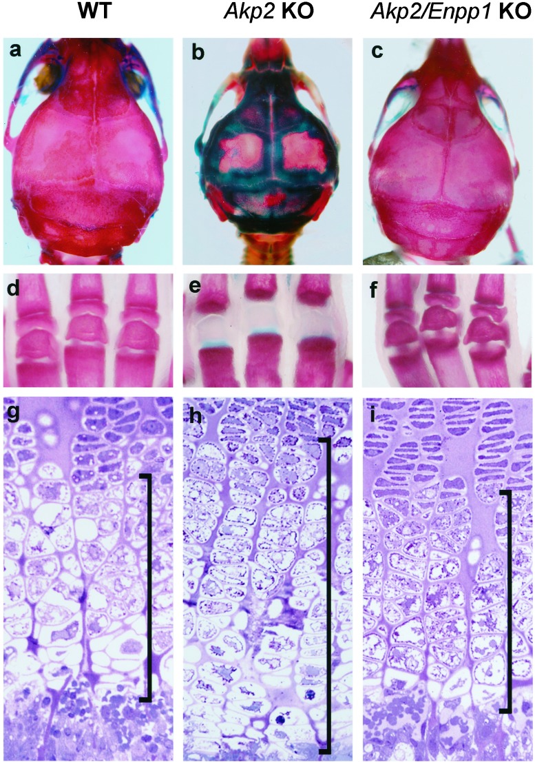
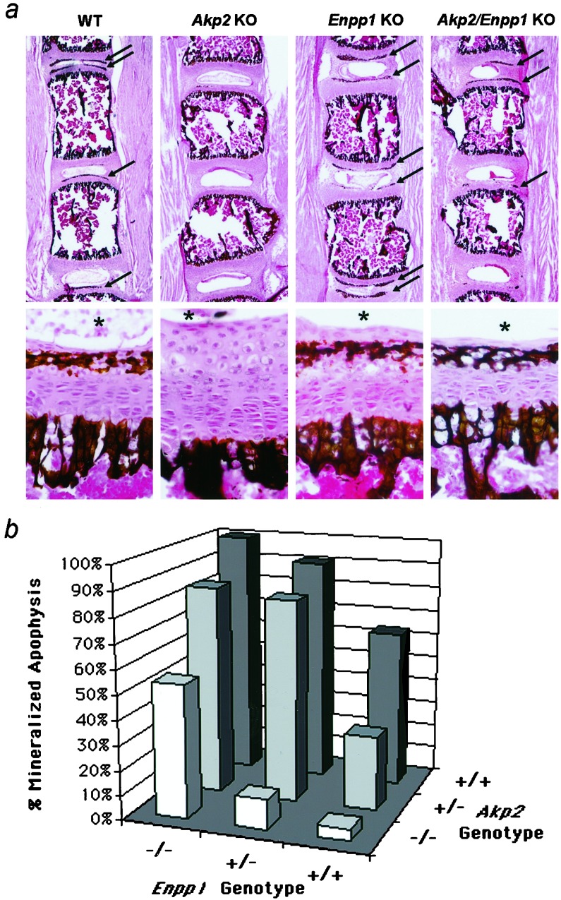
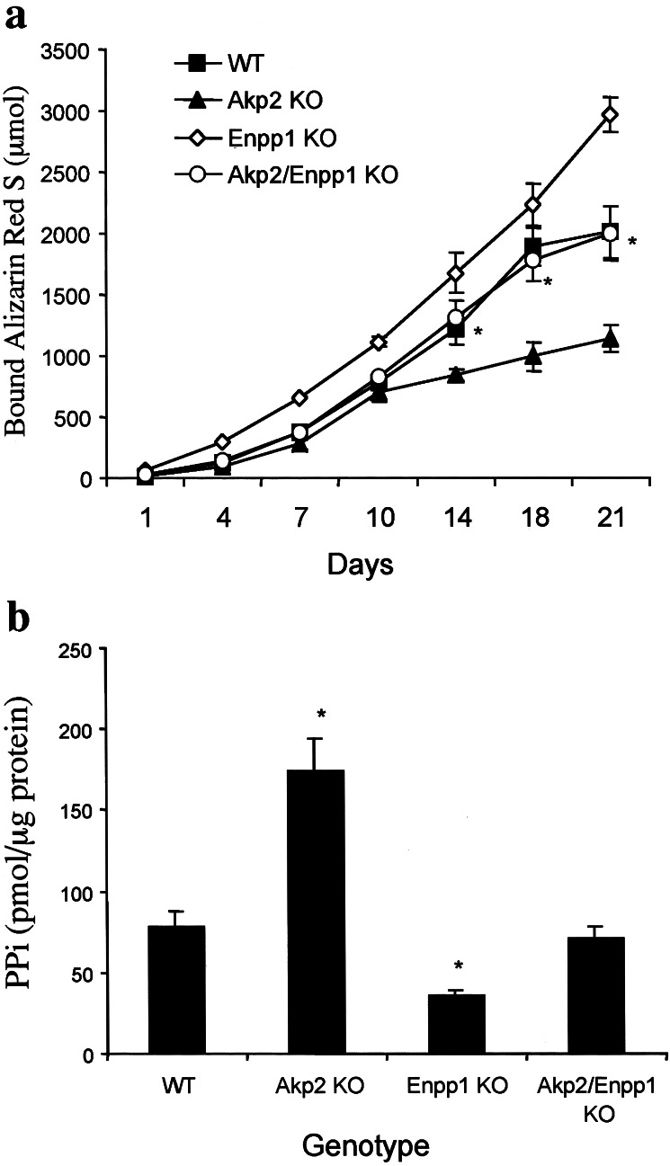
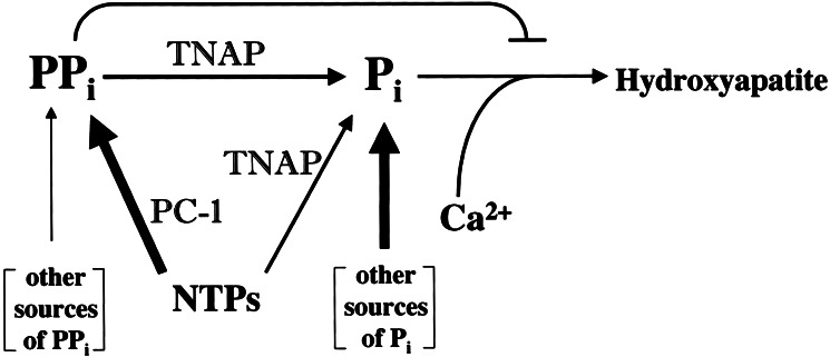
Osteoblasts mineralize bone matrix by promoting hydroxyapatite crystal formation and growth in the interior of membrane-limited matrix vesicles (MVs) and by propagating the crystals onto the collagenous extracellular matrix. Two osteoblast proteins, tissue-nonspecific alkaline phosphatase (TNAP) and plasma cell membrane glycoprotein-1 (PC-1) are involved in this process. Mutations in the TNAP gene result in the inborn error of metabolism known as hypophosphatasia, characterized by poorly mineralized bones, spontaneous fractures, and elevated extracellular concentrations of inorganic pyrophosphate (PP(i)). PP(i) suppresses the formation and growth of hydroxyapatite crystals. PP(i) is produced by the nucleoside triphosphate pyrophosphohydrolase activity of a family of isozymes, with PC-1 being the only member present in MVs. Mice with spontaneous mutations in the PC-1 gene have hypermineralization abnormalities that include osteoarthritis and ossification of the posterior longitudinal ligament of the spine. Here, we show the respective correction of bone mineralization abnormalities in knockout mice null for both the TNAP (Akp2) and PC-1 (Enpp1) genes. Each allele of Akp2 and Enpp1 has a measurable influence on mineralization status in vivo. Ex vivo experiments using cultured double-knockout osteoblasts and their MVs demonstrate normalization of PP(i) content and mineral deposition. Our data provide evidence that TNAP and PC-1 are key regulators of the extracellular PP(i) concentrations required for controlled bone mineralization. Our results suggest that inhibiting PC-1 function may be a viable therapeutic strategy for hypophosphatasia. Conversely, interfering with TNAP activity may correct pathological hyperossification because of PP(i) insufficiency.


Figures

Figure 1
Figure 1
Correction of skeletal mineralization and growth-plate structure in Akp2/Enpp1 double-KO mice. Whole-mount skeletal preparations of the calvaria (ac) and hind limb phalanges (df) show skeletal correction in Akp2/Enpp1 double-KO mice. The tissues were stained with Alizarin Red that detects bone mineral and Alcian Blue that stains unmineralized osteoid at pH 5.5. WT, calvaria, and phalanges of a 20-day-old WT mouse (a and d). At 3 weeks of age, the Enpp1 KO mice (not shown), do not show any evidence of hypermineralization in the calvaria or phalanges, and their whole-mount preparations are indistinguishable from those of WT mice. Akp2 KO, calvaria and phalanges of a 20-day-old Akp2 KO mouse (b and e). Note the absence of secondary ossification centers in the phalanges; Akp2/Enpp1 KO, calvaria and phalanges of a 20-day-old Akp2/Enpp1 double-KO mouse displaying correction of the mineralization defects (c and f). (gi) Nondecalcified sections of tibial growth plates of 10-day-old mice stained with hematoxylin/eosin. The WT tibial growth plate (g) shows normal proliferative zone cells at the top. Lightly stained hypertrophic zone cells are seen in the middle of the field, and the metaphysis is present at the bottom. In contrast, the tibial growth plate from Akp2 KO mice (h) shows a characteristic thickening, especially of the hypertrophic zone, as seen (23). Note the restoration of normal growth-plate thickness and cellularity in the tibial growth plate of Akp2/Enpp1 double-KO mice (i). Bars in the figure indicate the thickness of the hypertrophic zone of the growth plate. (Magnification ×310.)
Figure 2
Figure 2
Nondecalcified lumbar spine sections stained by the von Kossa procedure to visualize phosphate deposits. (a) Low (×16) and high (×240) magnification view of the 10-day-old vertebral apophyses of WT, Akp2 KO, Enpp1 KO, and Akp2/Enpp1 double-KO mice. Arrows point to mineralized secondary ossification centers in sample sections at low magnification. Note the absence of mineral deposits in sections from the Akp2 KO mice. *, relative position of the nucleus pulposus as a point of reference in the high magnification panels. (b) Graph plotting the contribution of each Akp2 and Enpp1 allele to the mineralization status of the lumbar vertebrae. The percentage of mineralized apophyses is plotted as a function of the Akp2 and Enpp1 genotypes.
Figure 3
Figure 3
Primary Akp2/Enpp1 double-KO osteoblasts show a correction in their mineralization capacity and normalized PPi concentrations in MVs. (a) Akp2 KO osteoblasts show poor mineralization after 21 days in culture, whereas the cultures of Enpp1 KO osteoblasts have a higher mineralization capacity compared with WT osteoblasts. Remarkably, the Akp2/Enpp1 double-KO and the WT cultures display indistinguishable mineralization properties (n = 7 mice each genotype, in replicates of 4). *, P < 0.05 compared with single gene KO osteoblasts. (b) The histogram displays the concentration of PPi in the MVs of each genotype. Akp2/Enpp1 double-KO MVs show PPi levels that are equivalent to those in WT vesicles. In contrast, MVs from single KO osteoblasts show either an elevation (Akp2 KO) or a reduction (Enpp1 KO) in their PPi concentration (n = 5 mice, in replicates of 3) relative to TTPP mice. *, P < 0.05 compared with WT MVs.
Figure 4
Figure 4
Schematic representation of the counterregulatory functions of TNAP and PC-1 in modulating extracellular PPi concentrations. (NTPs, nucleoside triphosphates). In the bone matrix, PC-1 is the major producer of PPi (thick arrows), which in turn has an inhibitory effect on hydroxyapatite deposition. TNAP has a positive influence on mineralization primarily by controlling the size of the inhibitory pool of PPi through its inorganic pyrophosphatase activity. TNAP also generates Pi by using NTPs and PPi as substrates, but other more major sources of Pi (thick arrow), e.g., intestinal absorption, are likely to contribute the bulk of the Pi needed for hydroxyapatite deposition.

References

    1. Anderson H C. Clin Orthop Relat Res. 1995;314:266–280. - PubMed
    1. Boskey A L. Connect Tissue Res. 1996;35:357–363. - PubMed
    1. Henthorn P, Millán J L, Leboy P. In: Principles of Bone Biology. Seibel M J, Robins S P, Bilezikian J P, editors. San Diego: Academic; 1999. pp. 127–136.
    1. Henthorn P S, Whyte M P. Clin Chem. 1992;38:2501–2505. - PubMed
    1. Whyte M P. In: The Metabolic and Molecular Bases of Inherited Disease. Scriver C R, Beaudet A L, Sly W S, Valle D, Childs B, Kinzler K W, Vogelstein B, editors. New York: McGraw–Hill; 2001. pp. 5313–5329.

Publication types

MeSH terms

Substances