. 2002 Jul-Aug;4(4):337-46.
doi: 10.1038/sj.neo.7900247.

Microarray and biochemical analysis of lovastatin-induced apoptosis of squamous cell carcinomas

Affiliations

Microarray and biochemical analysis of lovastatin-induced apoptosis of squamous cell carcinomas

Jim Dimitroulakos et al. Neoplasia. 2002 Jul-Aug.

Abstract

We recently identified 3-hydroxy-3-methylglutaryl coenzyme A (HMG-CoA) reductase, the rate-limiting enzyme of the mevalonate pathway, as a potential therapeutic target of the head and neck squamous cell carcinomas (HNSCC) and cervical carcinomas (CC). The products of this complex biochemical pathway, including de novo cholesterol, are vital for a variety of key cellular functions affecting membrane integrity, cell signaling, protein synthesis, and cell cycle progression. Lovastatin, a specific inhibitor of HMG-CoA reductase, induces a pronounced apoptotic response in a specific subset of tumor types, including HNSCC and CC. The mediators of this response are not well established. Identification of differentially expressed genes represents a feasible approach to delineate these mediators as lovastatin has the potential to modulate transcription indirectly by perturbing levels of sterols and other mevalonate metabolites. Expression analysis following treatment of the HNSCC cell lines SCC9 or SCC25 with 10 microM lovastatin for 1 day showed that less than 2% (9 cDNAs) of the 588 cDNAs on this microarray were affected in both cell lines. These included diazepam-binding inhibitor/acyl-CoA-binding protein, the activated transcription factor 4 and rhoA. Because the biosynthesis of mevalonate leads to its incorporation into more than a dozen classes of end products, their role in lovastatin-induced apoptosis was also evaluated. Addition of the metabolites of all the major branches of the mevalonate pathway indicated that only the nonsterol moiety, geranylgeranyl pyrophosphate (GGPP), significantly inhibited the apoptotic effects of lovastatin in HNSCC and CC cells. Because rhoA requires GGPP for its function, this links the microarray and biochemical data and identifies rhoA as a potential mediator of the anticancer properties of lovastatin. Our data suggest that the depletion of nonsterol mevalonate metabolites, particularly GGPP, can be potential mediators of lovastatin-induced apoptosis of HNSCC and CC cells.


Figures
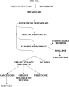
Figure 1
Figure 1
The mevalonate pathway.
Figure 2
Figure 2
(A) Differential expression analysis using the Clontech Atlas microarray between solvent control and treatment with 10 µM lovastatin in the SCC9 and SCC25 HNSCC-derived cell lines. (B) Differentially expressed genes were identified using the Atlas Image software. Of the 588 cDNAs represented on this slotblot, only nine demonstrated differential expression and are listed in the table with relative differences in expression (see Materials and Methods section for detailed description of analysis). The genes identified as potentially regulated by lovastatin included rhoA, extracellular signal-regulated kinase 1 (ERK1), insulin-like growth factor-binding protein 3 (IGFBP3), cdc25B, ATF-4, BTEB2, calgranulin, MIF-related protein, and DBI.
Figure 3
Figure 3
Promoter activity, as assessed by luciferase activity, of DBI and the ATF consensus constructs in SCC9 and SCC25. Cells were treated for 24 hours with solvent control or 10 µM lovastatin. Control levels of expression were normalized to one for ease of presentation.
Figure 4
Figure 4
(A) The effect of lovastatin on the expression of the HMG-CoA reductase (pRED-luc), the HMG-CoA synthase (pSYNTHASE-luc), and the thymidine kinase (pTK-luc) luciferase chimeric constructs in cos-7 cells. (B) Expression levels of pRED-luc, pSYNTHASE-luc, and pTK-luc in cos-7 cells constitutively expressing activated SREBP. Control levels of expression were normalized to one for ease of presentation.
Figure 5
Figure 5
(A) The effect of lovastatin on the expression of the DBI promoter, the DBI promoter with the SRE deleted, and the DBI promoter in cos-7 cells overexpressing activated SREBP. (B) The ATF consensus promoter luciferase chimeric construct in cos-7 cells and in cos-7 cells constitutively expressing activated SREBP. Cells were treated for 24 hours with solvent control or 10 µM lovastatin. Control levels of expression were normalized to one for ease of presentation.
Figure 6
Figure 6
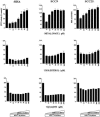
The effect of adding back mevalonate, cholesterol, or squalene on the cytotoxic effects of lovastatin in the CC cell line SIHA and the HNSCC cell lines SCC9 and SCC25. Cell viability was assessed using the MTT assay. Cultures were treated with solvent control, lovastatin at its predetermined LD50 (SIHA 15 µM; SCC9 50 µM; SCC25 35 µM), and increasing concentrations of mevalonate (top row), cholesterol (middle row), and squalene (bottom row) for 48 hours.
Figure 7
Figure 7
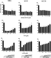
The effect of adding back ubiquinone, FPP, and GPP on the cytotoxic effects of lovastatin in the CC cell line SIHA and the HNSCC cell lines SCC9 and SCC25. Cell viability was assessed using the MTT assay. Cultures were treated with solvent control, lovastatin at its predetermined LD50 (SIHA 15 µM; SCC9 50 µM; SCC25 35 µM), and increasing concentrations of ubiquinone (top row), FPP (middle row), and GPP (bottom row) for 48 hours.
Figure 8
Figure 8
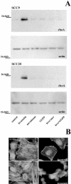
(A) Western blot analysis of rhoA and actin in SCC9 and SCC25 cells treated for 24 hours with either solvent control, 10 µM lovastatin, 100 µM mevalonate, 10 µM GGPP, and combinations of lovastatin and mevalonate or GGPP. (B) Immunofluorescence microscopy to examine the effect of lovastatin on actin cytoskeletal organization in SCC cells. FITC phalloidin staining of SCC9 and SIHA cells with solvent control or 10 µM lovastatin for 24 hours. Nuclei were counterstained with DAPI. Original magnification, x400.

Similar articles

Cited by

References

    1. Agarwal B, Rao CV, Bhendwal S, Ramey WR, Shirin H, Reddy BS, Holt PR. Lovastatin augments sulindac-induced apoptosis in colon cancer cells and potentiates chemopreventive effect of sulindac. Proc Am Assoc Cancer Res. 1999;40:57. - PubMed
    1. Aznar S, Lacal JC. Rho signals to cell growth and apoptosis. Cancer Lett. 2001;165:1–10. - PubMed
    1. Cooper CS. Applications of microarray technology in breast cancer research. Breast Cancer Res. 2001;3:158–175. - PMC - PubMed
    1. Corsini A, Maggi FM, Catapano AL. Pharmacology of competitive inhibitors of HMG-CoA reductase. Pharmacol Res. 1995;31:9–27. - PubMed
    1. Cunningham MJ. Genomics and proteomics: the new millennium of drug discovery and development. J Pharmacol Toxicol Methods. 2000;44:291–300. - PubMed

Publication types

MeSH terms

Substances