MRI-guided diffuse optical spectroscopy of malignant and benign breast lesions
- PMID: 12082551
- PMCID: PMC1661680
- DOI: 10.1038/sj.neo.7900244
MRI-guided diffuse optical spectroscopy of malignant and benign breast lesions
Abstract
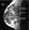
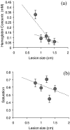
We present the clinical implementation of a novel hybrid system that combines magnetic resonance imaging (MRI) and near-infrared (NIR) optical measurements for the noninvasive study of breast cancer in vivo. Fourteen patients were studied with a MR-NIR prototype imager and spectrometer. A diffuse optical tomographic scheme employed the MR images as a priori information to implement an image-guided NIR localized spectroscopic scheme. All patients who entered the study also underwent gadolinium-enhanced MRI and biopsy so that the optical findings were cross-validated with MR readings and histopathology. The technique quantified the oxy- and deoxyhemoglobin of five malignant and nine benign breast lesions in vivo. Breast cancers were found with decreased oxygen saturation and higher blood concentration than most benign lesions. The average hemoglobin concentration ([H]) of cancers was 0.130+/-0.100 mM, and the average hemoglobin saturation (Y) was 60+/-9% compared to [H]=0.018+/-0.005 mM and Y=69+/-6% of background tissue. Fibroadenomas exhibited high hemoglobin concentration [H]=0.060+/-0.010 mM and mild decrease in oxygen saturation Y=67+/-2%. Cysts and other normal lesions were easily differentiated based on intrinsic contrast information. This novel optical technology can be a significant add-on in MR examinations and can be used to characterize functional parameters of cancers with diagnostic and treatment prognosis potential. It is foreseen that the technique can play a major role in functional activation studies of brain and muscle as well.
Figures


Similar articles
-
[Diagnostic value of preoperative contrast-enhanced MR imaging of the breast].Rofo. 2004 May;176(5):688-93. doi: 10.1055/s-2004-813119. Rofo. 2004. PMID: 15122467 German.
-
Fast 3D Near-infrared breast imaging using indocyanine green for detection and characterization of breast lesions.Rofo. 2011 Oct;183(10):956-63. doi: 10.1055/s-0031-1281726. Epub 2011 Oct 4. Rofo. 2011. PMID: 21972043
-
[Diagnostic value of ADC and rADC of diffusion weighted imaging in malignant breast lesions].Zhonghua Zhong Liu Za Zhi. 2010 Mar;32(3):217-20. Zhonghua Zhong Liu Za Zhi. 2010. PMID: 20450592 Chinese.
-
[Diagnostic immunohistochemistry of breast epithelial lesions].Ann Pathol. 2003 Dec;23(6):570-81. Ann Pathol. 2003. PMID: 15094595 Review. French.
-
[About mastopathy].Orv Hetil. 2007 Nov 25;148(47):2211-8. doi: 10.1556/OH.2007.28168. Orv Hetil. 2007. PMID: 18003579 Review. Hungarian.
Cited by
-
Optimization of image reconstruction for magnetic resonance imaging-guided near-infrared diffuse optical spectroscopy in breast.J Biomed Opt. 2015 May;20(5):56009. doi: 10.1117/1.JBO.20.5.056009. J Biomed Opt. 2015. PMID: 26000795 Free PMC article.
-
Computational Model for Tumor Oxygenation Applied to Clinical Data on Breast Tumor Hemoglobin Concentrations Suggests Vascular Dilatation and Compression.PLoS One. 2016 Aug 22;11(8):e0161267. doi: 10.1371/journal.pone.0161267. eCollection 2016. PLoS One. 2016. PMID: 27547939 Free PMC article.
-
Deep learning-based fusion of widefield diffuse optical tomography and micro-CT structural priors for accurate 3D reconstructions.Biomed Opt Express. 2023 Feb 7;14(3):1041-1053. doi: 10.1364/BOE.480091. eCollection 2023 Mar 1. Biomed Opt Express. 2023. PMID: 36950248 Free PMC article.
-
Machine learning model with physical constraints for diffuse optical tomography.Biomed Opt Express. 2021 Aug 23;12(9):5720-5735. doi: 10.1364/BOE.432786. eCollection 2021 Sep 1. Biomed Opt Express. 2021. PMID: 34692211 Free PMC article.
-
Optical imaging of the breast.Cancer Imaging. 2008 Nov 25;8(1):206-15. doi: 10.1102/1470-7330.2008.0032. Cancer Imaging. 2008. PMID: 19028613 Free PMC article.
References
-
- Chance B. Optical method. Annu Rev Biophys Biophys Chem. 1991;20:1–28. - PubMed
-
- Pogue BW, Poplack SP, McBride TO, Wells WA, Osterman KS, Osterberg UL, Paulsen KD. Quantitative hemoglobin tomography with diffuse near-infrared spectroscopy: pilot results in the breast. Radiology. 2001;218:261–266. - PubMed
-
- Benaron DA, Hintz SR, Villringer A, Boas D, Kleinschmidt A, Frahm J, Hirth C, Obrig H, van Houten JC, Kermit EL, Cheong WF, Stevenson DK. Noninvasive functional imaging of human brain using light. J Cereb Blood Flow Metab. 2000;20:469–477. - PubMed
Publication types
MeSH terms
Substances
Grants and funding
LinkOut - more resources
Full Text Sources
Other Literature Sources
Medical
Miscellaneous
