. 2002 Jul 9;99(14):9219-24.
doi: 10.1073/pnas.142053899. Epub 2002 Jun 25.

Interactions between Eph kinases and ephrins provide a mechanism to support platelet aggregation once cell-to-cell contact has occurred

Affiliations

Interactions between Eph kinases and ephrins provide a mechanism to support platelet aggregation once cell-to-cell contact has occurred

Nicolas Prevost et al. Proc Natl Acad Sci U S A. .

Abstract

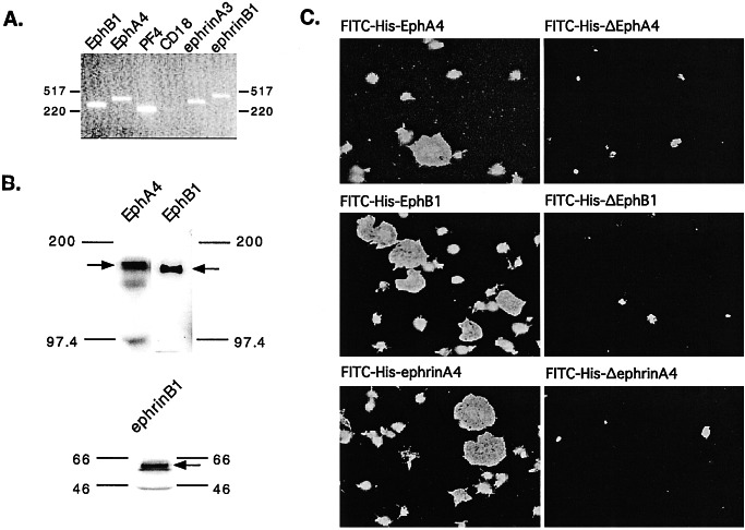
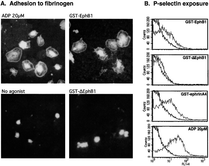
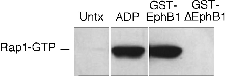
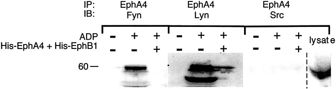
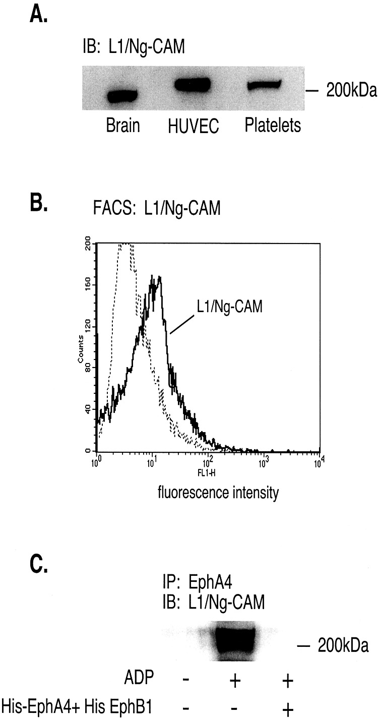
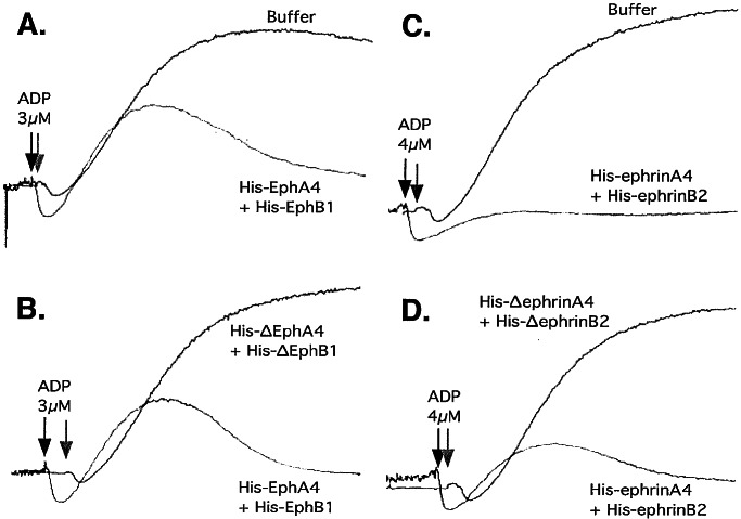
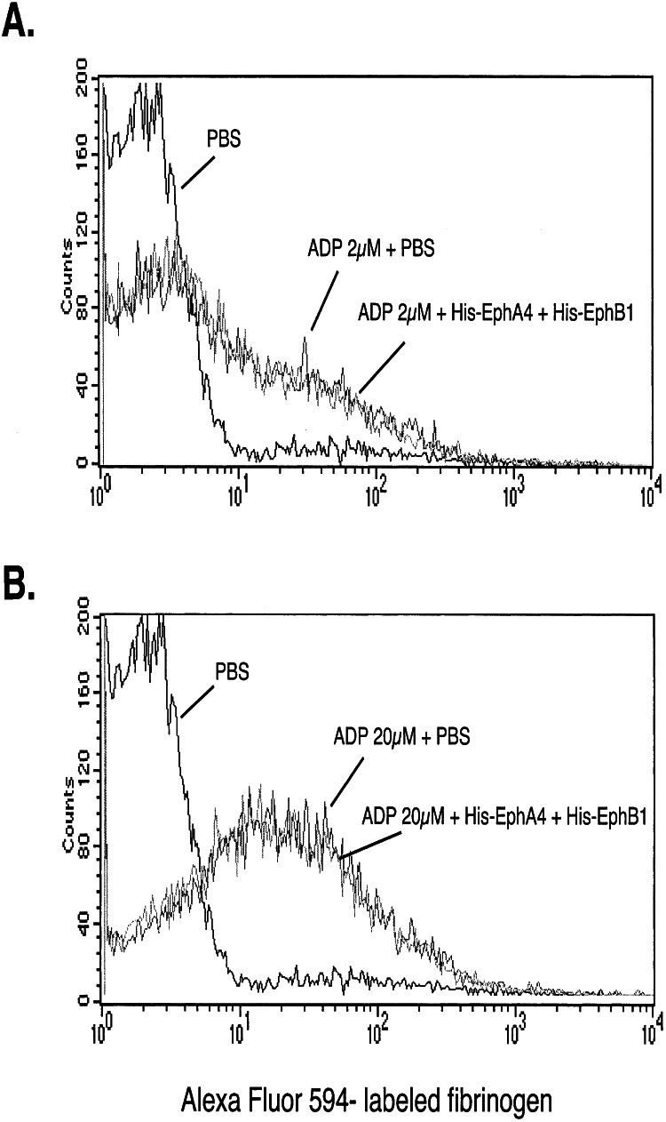
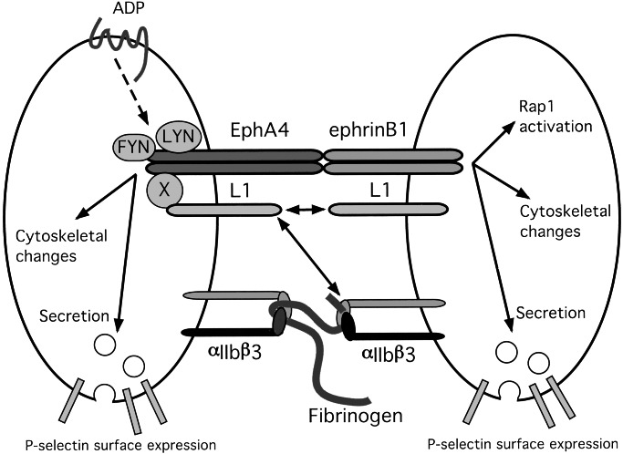
Eph kinases are receptor tyrosine kinases whose ligands, the ephrins, are also expressed on the surface of cells. Interactions between Eph kinases and ephrins on adjacent cells play a central role in neuronal patterning and vasculogenesis. Here we examine the expression of ephrins and Eph kinases on human blood platelets and explore their role in the formation of the hemostatic plug. The results show that human platelets express EphA4 and EphB1, and the ligand, ephrinB1. Forced clustering of EphA4 or ephrinB1 led to cytoskeletal reorganization, adhesion to fibrinogen, and alpha-granule secretion. Clustering of ephrinB1 also caused activation of the Ras family member, Rap1B. In platelets that had been activated by ADP and allowed to aggregate, EphA4 formed complexes with two tyrosine kinases, Fyn and Lyn, and the cell adhesion molecule, L1. Blockade of Eph/ephrin interactions prevented the formation of these complexes and caused platelet aggregation at low ADP concentrations to become more readily reversible. We propose that when sustained contacts between platelets have occurred in response to agonists such as collagen, ADP, and thrombin, the binding of ephrins to Eph kinases on adjacent platelets provides a mechanism to perpetuate signaling and promote stable platelet aggregation.


Figures

Figure 1
Figure 1
Evidence for Eph kinases and ephrins on the surface of human platelets. (A) Reverse transcription and amplification (RT-PCR) of platelet RNA using primers specific for EphB1, EphA4, ephrinA3, and ephrinB1. Platelet factor 4 (PF4) is specific for platelets, whereas CD18 was used as a marker for leukocyte contamination. Sequencing of the amplified products confirmed their identities. (B) Immunoblots of total platelet lysates with antibodies that recognize EphA4, EphB1, and ephrinB1. (C) Platelets were incubated with 100 nM phorbol 12-myristate 13-acetate, allowed to adhere and spread on a fibrinogen-coated surface, and then stained with FITC-conjugated, His-6-tagged recombinant proteins corresponding to the exodomains of EphA4, EphB1, and ephrinA4. Control proteins lacked residues required for Eph/ephrin interactions are denoted with a “Δ.”
Figure 2
Figure 2
Clustering of Eph kinases and ephrins activates platelets. (A) Platelets were incubated on a fibrinogen-coated surface for 20 min either without an agonist or with ADP (20 μM), GST-EphB1 (2 μg/ml), or GST-ΔEphB1 (2 μg/ml), and then stained with rhodamine-phalloidin to visualize actin. (B) Secretion of platelet α-granules was detected by flow cytometry by using a FITC-labeled antibody that recognizes P-selectin. The results shown compare resting with stimulated platelets and are representative of five such experiments.
Figure 3
Figure 3
Activation of Rap1B. Platelets were incubated for 5 min with ADP (20 μM), GST-EphB1 (10 μg/ml), or GST-ΔEphB1(10 μg/ml), after which Rap1B in the GTP-bound state was precipitated with GST-RBD and detected with an anti-Rap1 antibody. The results shown are representative of three such experiments.
Figure 4
Figure 4
Platelet aggregation results in the formation of Fyn and Lyn complexes with EphA4. Platelets were incubated with ADP (20 μM) under conditions that allowed aggregation to occur. After 5 min, the cells were lysed and EphA4 was immunoprecipitated. The precipitates were then subjected to electrophoresis and probed with antibodies to Fyn, Lyn, or Src. Where indicated, Eph/ephrin interactions were blocked with a combination of His-EphA4 plus His-EphB1 at a final concentration of 10 μg/ml of each, a concentration found to be sufficient to block ephrin/Eph kinase interactions (58, 59). The lane farthest to the right shows total platelet lysates probed with a Src-specific antibody to document that Src is present. IP, the immunoprecipitating antibody; IB, the antibody used in the Western immunoblot.
Figure 5
Figure 5
L1/Ng-CAM is expressed in human platelets and forms complexes with EphA4 during platelet aggregation. (A) Western blot of total cell lysates from human brain, endothelial cells (HUVEC) and platelets showing the presence of L1/Ng-CAM. (B) Fluorescence-activated cell sorter analysis of human platelets with an antibody directed against the L1 exodomain. (C) Precipitation of L1 with EphA4 by using an anti-EphA4 antibody. The lysates were prepared from platelets incubated with 20 μM ADP under aggregating conditions in the presence or absence of His-EphA4 plus His-EphB1 (10 μg/ml of each). IP, the immunoprecipitating antibody; IB, the antibody used in the Western immunoblot.
Figure 6
Figure 6
Blockade of Eph/ephrin interactions inhibits platelet aggregation. Washed platelets supplemented with Ca2+ and fibrinogen and stirred at 37°C were activated with ADP in the presence or absence of the His-tagged fusion proteins that are indicated in the figure (10 μg/ml of each). The results shown are representative of seven such experiments.
Figure 7
Figure 7
Blockade of Eph/ephrin interactions does not inhibit fibrinogen binding to αIIbβ3. The binding of labeled fibrinogen to platelets activated with ADP was measured by flow cytometry in the presence or absence of His-EphA4 plus His-EphB1 (10 μg/ml of each). The results shown compare resting with stimulated platelets and are representative of five such experiments.
Figure 8
Figure 8
A model for Eph/ephrin signaling in platelets. On the basis of the results of the present studies, it is proposed that when contacts between platelets become more than transient, the binding of ephrins on the surface of one platelet to cognate Eph kinases on adjacent platelets causes receptor clustering and promotes actin reorganization and α-granule secretion. Clustering of EphA4 in particular recruits Lyn, Fyn, and the cell adhesion molecule L1 (directly or through a potential intermediary protein X) into signaling complexes. In turn, that might affect the binding of L1, either with itself in a homophilic manner or with the β3 integrins, αIIbβ3, or αvβ3, although that has not yet been established.

Similar articles

Cited by

References

    1. Shattil S J. Thromb Haemostasis. 1999;82:318–325. - PubMed
    1. Angelillo-Scherrer A, De Frutos P G, Aparicio C, Melis E, Savi P, Lupu F, Arnout J, Dewerchin M, Hoylaerts M F, Herbert M, et al. Nat Med. 2001;7:215–221. - PubMed
    1. Wilkinson D G. Int Rev Cytol. 2000;196:177–244. - PubMed
    1. Dodelet V C, Pasquale E B. Oncogene. 2000;19:5614–5619. - PubMed
    1. Huynh-Do U, Stein E, Lane A A, Liu H, Cerretti D P, Daniel T O. EMBO J. 1999;18:2165–2173. - PMC - PubMed

Publication types

MeSH terms

Substances

LinkOut - more resources