. 2002 Jun;14(6):1391-403.
doi: 10.1105/tpc.000869.

The homologous ABI5 and EEL transcription factors function antagonistically to fine-tune gene expression during late embryogenesis

Affiliations

The homologous ABI5 and EEL transcription factors function antagonistically to fine-tune gene expression during late embryogenesis

Sandra Bensmihen et al. Plant Cell. 2002 Jun.

Abstract

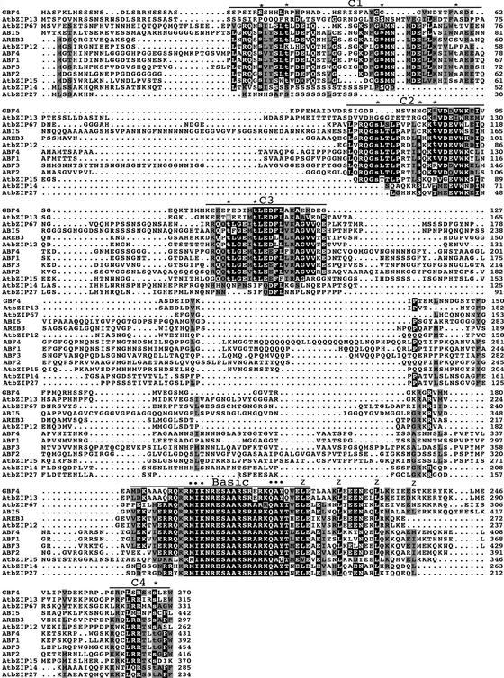
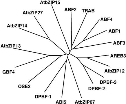
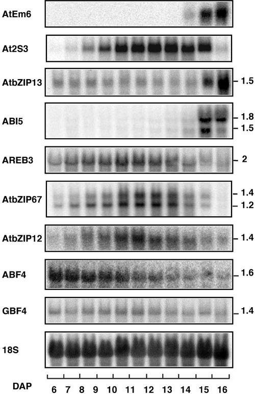
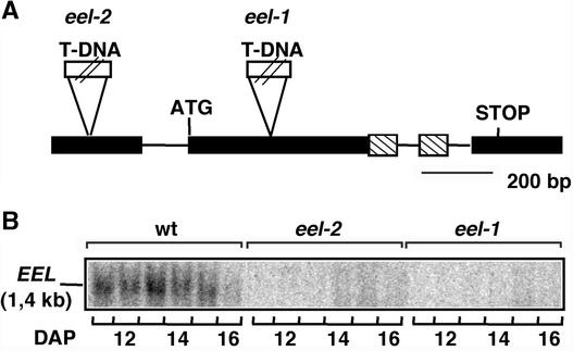
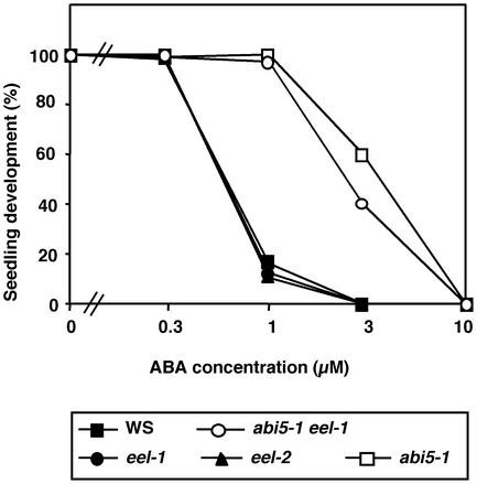
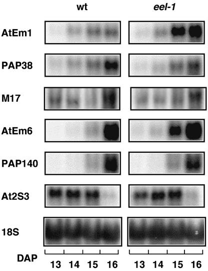
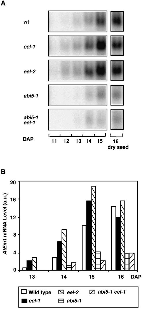
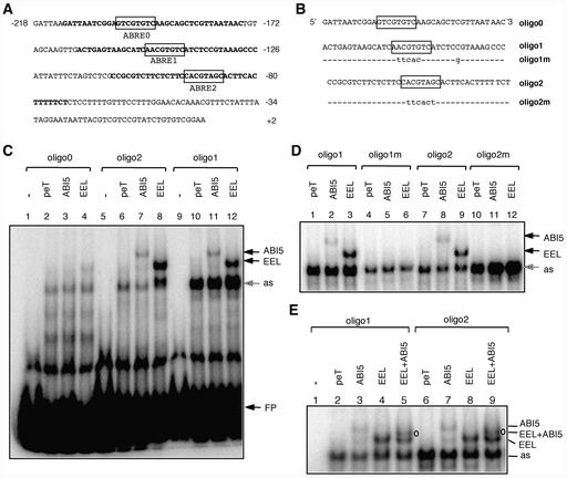
In Arabidopsis, the basic leucine zipper transcription factor ABI5 activates several late embryogenesis-abundant genes, including AtEm1 and AtEm6. However, the expression of many other seed maturation genes is independent of ABI5. We investigated the possibility that ABI5 homologs also participate in the regulation of gene expression during seed maturation. We identified 13 ABI5-related genes in the Arabidopsis genomic sequence. RNA gel blot analysis showed that seven of these genes are active during seed maturation and that they display distinct expression kinetics. We isolated and characterized two mutant alleles of one of these genes, AtbZIP12/EEL. Unlike abi5, the eel mutations did not inhibit the expression of any of the maturation marker genes that we monitored. On the contrary, the accumulation of the AtEm1 and AtEm6 mRNAs was enhanced in eel mutant seeds compared with wild-type seeds. Gel mobility shift assays, combined with analysis of the genetic interactions among the eel and abi5 mutations, indicated that ABI5 and EEL compete for the same binding sites within the AtEm1 promoter. This study illustrates how two homologous transcription factors can play antagonistic roles to fine-tune gene expression.


Figures

Figure 1.
Figure 1.
Sequence Comparison of Arabidopsis bZIP Proteins of the ABI5/ABF/AREB Family. Alignment of the predicted amino acid sequences of the 13 members of the ABI5/ABF/AREB family. Identical and conserved residues are shaded in black and gray, respectively. The positions of the C1 to C4 conserved domains and of the basic region are indicated by lines above the protein sequences. Potential phosphoresidues corresponding to consensus phosphorylation sites for Ca2+-dependent protein kinase (R/K-X-X-S/T), protein kinase C (S/T-X-K/R), cGMP-dependent protein kinase (K/R-X-X-X-S/T), or casein kinase II (S/T-X-X-D/E) are shown in lowercase letters, and their positions are indicated by asterisks. Residues from the basic region specific to this subfamily of bZIP are indicated by dots. The positions of the conserved Leu residues in the Leu zipper domain are indicated by z's. Protein sequence alignments were performed using ClustalW at the European Bioinformatics Institute (http://www.ebi.ac.uk/clustalw/) and edited using Macboxshade 2.11 software (Institute of Animal Health, Pirbright, Surrey, UK).
Figure 2.
Figure 2.
Relation Tree of bZIP Proteins from Arabidopsis and Other Plant Species. This tree includes the 13 Arabidopsis proteins shown in Figure 1, DPBF-1, DPBF-2, and DPBF-3 from sunflower, and TRAB1 and OSE2 from rice. Proteins were aligned using ClustalW, and the tree was constructed using Treeview software.
Figure 3.
Figure 3.
RNA Gel Blot Analysis of Gene Expression during Silique Development. Each of the indicated probes was hybridized to 12 μg of total RNA isolated from Wassilewskija (Ws) wild-type siliques harvested between 6 and 16 DAP. An 18S rRNA probe was used as a control for equal amounts of RNA loading in the different lanes. Estimated bZIP transcript sizes (kb) are indicated at right.
Figure 4.
Figure 4.
Insertion Mutants in AtbZIP12/EEL. (A) Structure of the AtbZIP12/EEL gene. Lines indicate introns, and boxes represent exons. The gene region encoding the basic region of the EEL protein is indicated by hatched boxes. Positions of the T-DNA insertions in the eel mutants are shown. (B) RNA gel blot analysis of EEL expression in the Ws wild type (wt) and eel-1 and eel-2 mutants. The EEL-specific probe was hybridized to 12 μg of total RNA from siliques of the indicated genotypes harvested between 11 and 16 DAP.
Figure 5.
Figure 5.
ABA Inhibition of Seedling Establishment. Seeds of Ws wild type (closed squares), eel-1 (closed circles), eel-2 (closed triangles), abi5-1 (open squares), and abi5-1 eel-1 (open circles) were plated on medium supplemented with the indicated concentrations of ABA, chilled for 4 days at 4°C in darkness, and incubated for 3 days at 21°C with a 16-h photoperiod. The number of seedlings with green cotyledons is expressed as the percentage of the total number of seeds plated (40 to 280 seeds).
Figure 6.
Figure 6.
Comparative Gel Blot Analysis of the Expression of Marker mRNAs during Wild-Type and eel-1 Silique Development. The indicated gene-specific probes were hybridized to 12 μg of total RNA from siliques of the Ws wild type (wt) and the eel-1 mutant harvested between 13 and 16 DAP. An 18S rRNA probe was used as a control for equal amounts of RNA loading in the different lanes.
Figure 7.
Figure 7.
Gel Blot Analysis of AtEm1 mRNA Expression during Silique Development. (A) Developmental time course of AtEm1 expression in the Ws wild type (wt), the eel-1, eel-2, and abi5-1 mutants, and the abi5-1 eel-1 double mutant. The AtEm1 probe was hybridized to 12 μg of total RNA from siliques of the indicated genotypes harvested between 11 and 16 DAP. (B) Levels of AtEm1 mRNA. Hybridization signals shown in (A) were quantified using a Storm imager and were normalized using the signals obtained after stripping and hybridizing the blot with an 18S rRNA probe. AtEm1 mRNA levels are expressed in arbitrary units (a.u.).
Figure 8.
Figure 8.
In Situ Hybridization Analysis of AtEm1 expression. In situ hybridization was performed on sections of Ws (A), abi5-1 (B), and eel-1 ([C] and [D]) embryos with an AtEm1-specific probe. In Ws and eel-1, the AtEm1 transcript (brown signal) was detected primarily in the provascular tissues (pv), the root tip (rt), and the shoot apical meristem (sam). In abi5-1 embryos, the AtEm1 signal was either absent or of reduced intensity. Embryos were harvested at 14 to 15 DAP. Bars = 50 μm.
Figure 9.
Figure 9.
ABI5 and EEL Interact with the Same ABREs in the AtEm1 Promoter. (A) Sequence of the minimal AtEm1 promoter. Nucleotides are numbered relative to the transcription start site (+1). ABRE motifs are shown in boxes. Promoter regions used as probes for EMSA are indicated in boldface. (B) Sequences of the wild-type (oligo0, oligo1, and oligo2) and mutated (oligo1m and oligo2m) oligonucleotides used as probes for EMSA. Mutated nucleotides are indicated in lowercase letters. Sequences are presented in the 5′ to 3′ orientation and are shown as single strand only. (C) ABI5 and EEL bind to oligo1 and oligo2. Oligo0 (lanes 1 to 4), oligo2 (lanes 5 to 8), and oligo1 (lanes 9 to 12) were incubated without protein extract (free probe; lanes 1, 5, and 9) or with in vitro–transcribed and translated vector pET16b (negative control; lanes 2, 6, and 10), ABI5 (lanes 3, 7, and 11), or EEL (lanes 4, 8, and 12). Bands shifted by ABI5 or EEL are indicated by arrows. as, an aspecific band shifted by reticulocyte proteins; FP, free probe. (D) Mutations in ABRE1 and ABRE2 abolish the binding of ABI5 and EEL. Oligo1 (lanes 1 to 3), oligo1m (lanes 4 to 6), oligo2 (lanes 7 to 9), or oligo2m (lanes 10 to 12) were incubated with in vitro–transcribed and translated vector pET16b (negative control; lanes 1, 4, 7, and 10), ABI5 (lanes 2, 5, 8, and 11), or EEL (lanes 3, 6, 9, and 12). In contrast to the wild-type oligo1 and oligo2, the mutant oligo1m and oligo2m were not shifted by ABI5 and EEL proteins. (E) ABI5 and EEL bind to oligo1 and oligo2 as homodimers and as a heterodimer. Oligo1 (lanes 1 to 5) or oligo2 (lanes 6 to 9) were incubated without protein extract (free probe; lane 1) or with in vitro–transcribed and translated vector pET16b (negative control; lanes 2 and 6), ABI5 (lanes 3 and 7), EEL (lanes 4 and 8), or cotranslated ABI5 and EEL (lanes 5 and 9). The shifted band corresponding to the ABI5-EEL heterodimer is indicated (EEL + ABI5).

References

    1. Asamizu, E., Nakamura, Y., Sato, S., and Tabata, S. (2000). A large scale analysis of cDNA in Arabidopsis thaliana: Generation of 12,028 non-redundant expressed sequence tags from normalized and size-selected cDNA libraries. DNA Res. 7, 175–180. - PubMed
    1. Bechtold, N., Ellis, J., and Pelletier, G. (1993). In planta Agrobacterium mediated gene transfer by infiltration of adult Arabidopsis thaliana plants. C. R. Acad. Sci. 316, 1194–1199. - PubMed
    1. Chern, M.S., Bobb, A.J., and Bustos, M.M. (1996). The regulator of MAT2 (ROM2) protein binds to early maturation promoters and represses PvALF-activated transcription. Plant Cell 8, 305–321. - PMC - PubMed
    1. Choi, H., Hong, J., Ha, J., Kang, J., and Kim, S.Y. (2000). ABFs, a family of ABA-responsive element binding factors. J. Biol. Chem. 275, 1723–1730. - PubMed
    1. Delseny, M., Bies-Etheve, N., Carles, C., Hull, G., Vicient, C.M., Raynal, M., Grellet, F., and Aspart, L. (2001). Late embryogenesis abundant (LEA) protein gene regulation during Arabidopsis seed maturation. J. Plant Physiol. 158, 419–427.

Publication types

MeSH terms