. 2002 Jun 25;99(13):8524-9.
doi: 10.1073/pnas.142224499.

Specificity and affinity motifs for Grb2 SH2-ligand interactions

Affiliations

Specificity and affinity motifs for Grb2 SH2-ligand interactions

Helmut W H G Kessels et al. Proc Natl Acad Sci U S A. .

Abstract

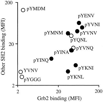
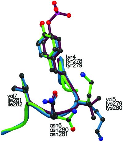
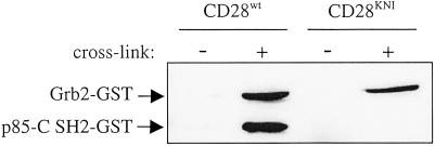
Protein-protein interactions are often mediated by the recognition of short continuous amino acid stretches on target proteins by specific binding domains. Affinity-based selection strategies have successfully been used to define recognition motifs for a large series of such protein domains. However, in many biological systems specificity of interaction may be of equal or greater importance than affinity. To address this issue we have developed a peptide library screening technology that can be used to directly define ligands for protein domains based on both affinity and specificity of interaction. We demonstrate the value of this approach by the selection of peptide ligands that are either highly specific for the Grb2 Src homology 2 (SH2) domain or that are cross-reactive between a group of related SH2 domains. Examination of previously identified physiological ligands for the Grb2 SH2 domain suggests that for these ligands regulation of the specificity of interaction may be an important factor for in vivo ligand selection.


Figures

Figure 1
Figure 1
Selection of peptides that bind the anti-β-endorphin (3-E7) mAb. A degenerate octa-peptide library was screened for library beads that bind 3-E7 antibody, and it was analyzed by flow cytometry. The percentage of fluorochrome-labeled beads was determined for all amino acids on each position. From left to right, screening for first, second, third, and fourth positions.
Figure 2
Figure 2
Amino acids at the first position C-terminal from the phospho-tyrosine determine Grb2 SH2 specificity. ASpYEXXXSA (A and C) and ASpYKXXXSA (B and D) phospho-peptide sublibraries were stained with FITC-labeled Grb2-GST versus either PE-labeled Grb2(R67H)-GST (A and B) or PE-labeled Grb2(R67H)-GST, Abl, Nck, PI3Kp85-N, PI3Kp85-C, SHP-2N, SHP-2C, and Src SH2-GST fusion proteins (C and D). Sublibraries were analyzed by flow cytometry. Results are represented as flow cytometry dot plots where each bead is represented by a dot. The percentage of Grb2-specific sequences within each library was determined as the fraction of dots that fall within the quadrangle shown in the lower right corner of the plots.
Figure 3
Figure 3
Selection of affinity and specificity motifs for the Grb2 SH2 domain. A degenerate phospho-tyrosine library was screened for Grb2 SH2-specific sequences and analyzed by flow cytometry. Libraries were screened for Grb2 SH2 optimal binding motifs (AC) and Grb2 SH2 specificity motifs (DF). A/D, B/E, and C/F represent screenings for amino acids at p+1, p+2, and p+3, respectively. The percentages of Grb2 SH2-specific sequences were determined as the percentage of gated beads as shown in Fig. 2.
Figure 4
Figure 4
Binding affinity of phospho-peptide motifs for Grb2. The capacity of the indicated soluble phospho-peptides to compete with horseradish peroxidase-conjugated ASpYVNVSA tetramers for binding to Grb2-GST was measured by ELISA. Competitor peptides used were: ASpYQNLSA (■), ASpYKNISA (●), ASpYVNVSA (□), and the Src SH2-specific sequence ASpYEEISA (○). Data shown are means of quadruplicates ± SD.
Figure 5
Figure 5
Specificity of Grb2 SH2 binding to naturally occurring ligands. A set of naturally occurring binding motifs was synthesized onto beads and analyzed for their capacities to bind Grb2-GST (0.1 μM, x axis) or a panel of other SH2 domains (1.0 μM each, y axis) by measuring the mean fluorescence intensity (MFI) by flow cytometry. ●, Beads that contain naturally occurring Grb2-binding motifs. ○, Non-naturally occurring Grb2-binding motifs pYQNL and pYVNQ, and the p85-specific pYMDM, pYGGG and unphosphorylated YVNV as negative controls.
Figure 6
Figure 6
YKNI sites in SHP-1 and SHP-2 display a β-turn conformation similar to a Grb2-bound phospho-peptide ligand. The YKNI (278–281) loop from SHP-1 (blue) and the YKNI (279–282) loop from SHP-2 (green) are overlaid with the structure of a Grb2 SH2-bound phospho-peptide ligand pYVNV (purple) (37). The rms differences between Cα backbones of SHP-1 loop and Grb2 ligand and SHP-2 and Grb2 ligand are 0.286 Å and 0.134 Å, respectively.
Figure 7
Figure 7
CD28KNI recruited Grb2 but lost the ability to bind PI3Kp85-C SH2. Jurkat T cells expressing either CD28wt or CD28KNI were activated with anti-CD28 mAbs or left unstimulated. CD28 was immunoprecipitated in the presence of soluble biotinylated Grb2-GST and PI3Kp85-C SH2-GST fusion proteins, and association of these proteins with CD28 was revealed by Western blot analysis.

Similar articles

Cited by

References

    1. Sudol M. Oncogene. 1998;17:1469–1474. - PubMed
    1. Anderson D, Koch C A, Grey L, Ellis C, Moran M F, Pawson T. Science. 1990;250:979–982. - PubMed
    1. Songyang Z, Shoelson S E, Chaudhuri M, Gish G, Pawson T, Haser W G, King F, Roberts T, Ratnofsky S, Lechleider R J, et al. Cell. 1993;72:767–778. - PubMed
    1. Songyang Z, Shoelson S E, McGlade J, Olivier P, Pawson T, Bustelo X R, Barbacid M, Sabe H, Hanafusa H, Yi T, et al. Mol Cell Biol. 1994;14:2777–2785. - PMC - PubMed
    1. Oligino L, Lung F D, Sastry L, Bigelow J, Cao T, Curran M, Burke T R, Jr, Wang S, Krag D, Roller P P, et al. J Biol Chem. 1997;272:29046–29052. - PubMed

Publication types