. 2002 Jun 25;99(13):8862-7.
doi: 10.1073/pnas.142078099.

Urokinase-urokinase receptor interaction mediates an inhibitory signal for HIV-1 replication

Affiliations

Urokinase-urokinase receptor interaction mediates an inhibitory signal for HIV-1 replication

Massimo Alfano et al. Proc Natl Acad Sci U S A. .

Abstract

Elevated levels of soluble urokinase-type plasminogen activator (uPA) receptor, CD87/u-PAR, predict survival in individuals infected with HIV-1. Here, we report that pro-uPA (or uPA) inhibits HIV-1 expression in U937-derived chronically infected promonocytic U1 cells stimulated with phorbol 12-myristate 13-acetate (PMA) or tumor necrosis factor-alpha (TNF-alpha). However, pro-uPA did not inhibit PMA or TNF-alpha-dependent activation of nuclear factor-kB or activation protein-1 in U1 cells. Cell-associated HIV protein synthesis also was not decreased by pro-uPA, although the release of virion-associated reverse transcriptase activity was substantially inhibited, suggesting a functional analogy between pro-uPA and the antiviral effects of IFNs. Indeed, cell disruption reversed the inhibitory effect of pro-uPA on activated U1 cells, and ultrastructural analysis confirmed that virions were preferentially retained within cell vacuoles in pro-uPA treated cells. Neither expression of endogenous IFNs nor activation of the IFN-inducible Janus kinase/signal transducer and activator of transcription pathway were induced by pro-uPA. Pro-uPA also inhibited acute HIV replication in monocyte-derived macrophages and activated peripheral blood mononuclear cells, although with great inter-donor variability. However, pro-uPA inhibited HIV replication in acutely infected promonocytic U937 cells and in ex vivo cultures of lymphoid tissue infected in vitro. Because these effects occurred at concentrations substantially lower than those affecting thrombolysis, pro-uPA may represent a previously uncharacterized class of antiviral agents mimicking IFNs in their inhibitory effects on HIV expression and replication.


Figures
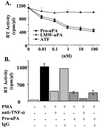
Figure 1
Figure 1
Pro-uPA and ATF, but not LMW-uPA, inhibit HIV-1 expression in PMA-stimulated U1 cells. Additive suppression by pro-uPA and anti-TNF-α Ab. (A) U1 cells were stimulated with PMA in the presence or absence of pro-uPA, LMW-uPA, or ATF; values represent the peak levels of RT activity achieved after 72 h of stimulation. A similar pattern of inhibition was observed after 6 days of stimulation (not shown). (B) Anti-TNF-α Ab (1 μg/ml), isotype Ab, and pro-uPA (10 nM) were added, either alone or in combination, 20 min before PMA stimulation of U1 cells. The results represent the means ± SD of three independent experiments.
Figure 2
Figure 2
Pro-uPA inhibits HIV-1 expression but not NF-kB activation induced by TNF-α or PMA stimulation of U1 cells. (A) U1 cells were preincubated for 20 min with different concentrations of pro-uPA and then stimulated with TNF-α (1 ng/ml). The results represent peak RT activity observed after 72 h of stimulation. (B) WCE prepared 30 min and 4 h after PMA or TNF-α stimulation of U1 cells were analyzed for NF-kB activation. U1 cells express a banding pattern typical of minus U937 cell clones, in that truncated p65 heterodimerizes with p50, as reported (16). The last two lanes on the right indicate supershifting of the bound complexes by anti-p50 and anti-p65 Abs.
Figure 3
Figure 3
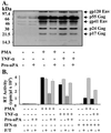
Posttranslational inhibition of HIV-1 expression from U1 cells by pro-uPA. (A) HIV-1 protein synthesis was analyzed by Western blotting 20 h after stimulation in the presence or absence of pro-uPA. Molecular weight markers and the main viral proteins are indicated on the left and on the right, respectively. (B) Reversion of pro-uPA (10 nM) mediated inhibition of HIV expression in stimulated U1 cells by cell disruption. F/T indicates that the cells were disrupted by five cycles of freezing and thawing.
Figure 4
Figure 4
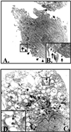
Preferential accumulation of HIV-1 virions in intracytoplasmatic vacuoles in PMA-stimulated U1 cells in the presence of pro-uPA. U1 cells were stimulated for 48 h with (A) PMA (magnification ×11,000) or (B) PMA plus pro-uPA (10 nM) (magnification ×15,000). (B and D) Enlargement of a detail of A and C, respectively. Two independent experiments were performed with similar results. Both unstimulated and pro-uPA-treated U1 cells did not show evidence of virion expression, whereas approximately 50% of PMA-stimulated cells were positive for virion expression, as reported (31, 32).
Figure 5
Figure 5
Pro-uPA inhibits HIV-1 replication in U937 plus and minus cells. (A) Constitutive expression of uPAR and CD11b/CD18 in U937 cells. Cells were identified based on their light scatter and analyzed for markers expression. Isotype analysis is shown as black histograms. Minus U937 cells constitutively express higher levels of uPAR (bold line) and CD11b/CD18 (dotted line) than plus cells. (B) U937 cell clones were pretreated with pro-uPA (1 nM) and infected with the X4 HIV-1LAI/IIIB virus. Fresh medium supplemented with pro-uPA (1 nM) was added to the cell cultures every 72 h.
Figure 6
Figure 6
Pro-uPA inhibits HIV-1 replication in lymphoid tissues. Pro-uPA (10 nM) was added either 20 min before or after infection of lymphoid tissue by the R5 HIV-1BaL. Data are shown as a mean ± SD of six independent cultures from one representative of three independent experiments performed.

References

    1. Dano K, Andreasen P A, Grondahl-Hansen J, Kristensen P, Nielsen L S, Skriver L. Adv Cancer Res. 1985;44:139–266. - PubMed
    1. Blasi F, Vassalli J D, Dano K. J Cell Biol. 1987;104:801–804. - PMC - PubMed
    1. Plesner T, Behrendt N, Ploug M. Stem Cells. 1997;15:398–408. - PubMed
    1. Gyetko M R, Libre E A, Fuller J A, Chen G H, Toews G. J Lab Clin Med. 1999;133:274–288. - PubMed
    1. May A E, Kanse S M, Lund L R, Gisler R H, Imhof B A, Preissner K T. J Exp Med. 1998;188:1029–1037. - PMC - PubMed

Publication types

MeSH terms

Substances

LinkOut - more resources