. 2002 Jun 25;99(13):8874-9.
doi: 10.1073/pnas.132269299. Epub 2002 Jun 19.

Stable expression of calpain 3 from a muscle transgene in vivo: immature muscle in transgenic mice suggests a role for calpain 3 in muscle maturation

Affiliations

Stable expression of calpain 3 from a muscle transgene in vivo: immature muscle in transgenic mice suggests a role for calpain 3 in muscle maturation

M J Spencer et al. Proc Natl Acad Sci U S A. .

Abstract

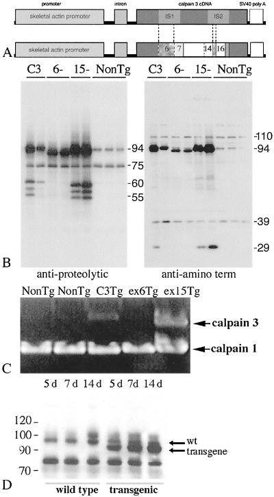
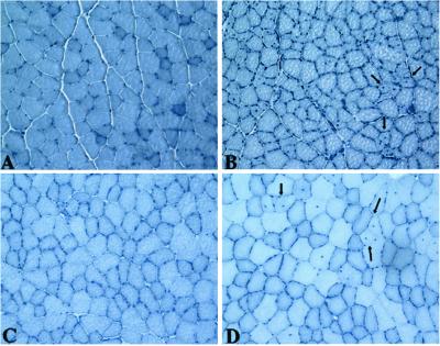
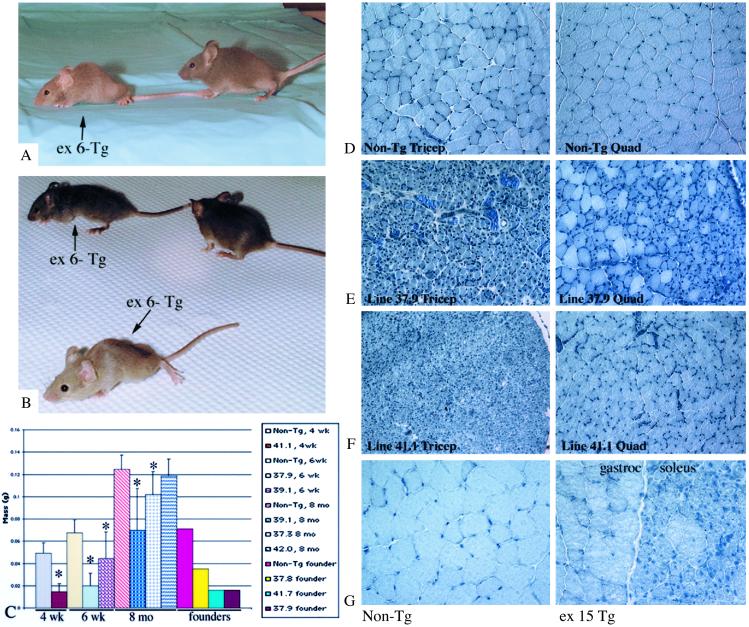
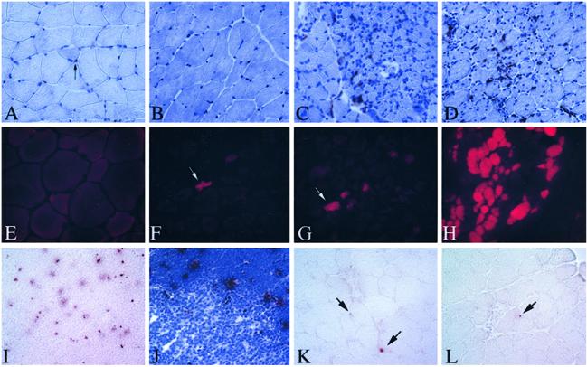
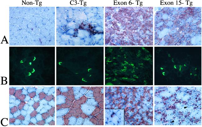
Limb-girdle muscular dystrophy, type 2A (LGMD 2A), is an autosomal recessive disorder that causes late-onset muscle-wasting, and is due to mutations in the muscle-specific protease calpain 3 (C3). Although LGMD 2A would be a feasible candidate for gene therapy, the reported instability of C3 in vitro raised questions about the potential of obtaining a stable, high-level expression of C3 from a transgene in vivo. We have generated transgenic (Tg) mice with muscle-specific overexpression of full-length C3 or C3 isoforms, which arise from alternative splicing, to test whether stable expression of C3 transgenes could occur in vivo. Unexpectedly, we found that full-length C3 can be overexpressed at high levels in vivo, without toxicity. In addition, we found that Tg expressing C3 lacking exon 6, an isoform expressed embryonically, have muscles that resemble regenerating or developing muscle. Tg expressing C3 lacking exon 15 shared this morphology in the soleus, but not other muscles. Assays of inflammation or muscle membrane damage indicated that the Tg muscles were not degenerative, suggesting that the immature muscle resulted from a developmental block rather than degeneration and regeneration. These studies show that C3 can be expressed stably in vivo from a transgene, and indicate that alternatively spliced C3 isoforms should not be used in gene-therapy applications because they impair proper muscle development.


Figures

Figure 1
Figure 1
Tg constructs, protein expression, and analysis of transgene activity. (A) Constructs used to make Tg mice. Ex deletions are indicated by dotted lines. Numbers are ex numbers. (B) Immunoblot of muscle extracts from Tgs probed with 12A2 (left blot) or pNS (right blot). Wild-type C3 is 94 kDa. Deletion isoforms produce 89.5-kDa (ex 6) and 93-kDa (ex 15) proteins. (C) Zymogram of Tg muscle extracts. (D) Time course of transgene expression. Whole-muscle extracts from 5-, 7-, and 14-day (d) non-Tg and ex 6− Tg were immunoblotted for C3. Arrows indicate wt and transgene products.
Figure 2
Figure 2
Morphology of C3 Tg Muscles. (AD) Cross sections of muscles stained with hematoxylin. (A) Non-Tg 6-week quad. (B) Line 7.9, 6-week quad. (C) Line 7.2, 16-week quad. (D) Line 7.2, 16-week tibialis anterior. Arrows point to centrally located nuclei. All micrographs were photographed at the same magnification.
Figure 3
Figure 3
Phenotype of ex 6− and ex 15− Tg. (A and B) Photographs of ex 6− Tg and non-Tg littermates. Arrows indicate ex 6− Tg mice. Non-Tg littermates do not have arrows. (C) Graph represents average muscle masses of 4-week, 6-week, and 8-month triceps muscles of different ex 6− Tg lines. *, P < 0.05 is significantly different from age-matched controls. Non-Tg controls are the left-most bar for each age group. Weights of founder triceps also are shown on the graph. For each of these founder bars, n = 1. All others are n > 5, except for 37.9, which is n = 2. Vertical bars represent SD. (DF) Sections of 4- to 6-week non-Tg and ex 6− Tg muscles stained with hematoxylin. All ex 6− micrographs were photographed at the same magnification. (G) Sections of non-Tg gastrocnemius and ex 15− Tg gastrocnemius/soleus. Both micrographs were taken at the same magnification.
Figure 4
Figure 4
Absence of indicators of necrosis/degeneration in Tg mice. (AD) Sections of Tg soleus stained with mac 1 (red) and counterstained with hematoxylin (blue). (A) Non-Tg. (B) C3-Tg. (C) Ex 6− Tg. (D) Ex 15− Tg. (EH) Triceps muscles from EB-injected mice (E) non-Tg mouse, (F and G) ex 6− Tg, (H) quadriceps from an mdx mouse. EB shows red fluorescence under UV light. (IL) TUNEL-stained sections. (I and J) TUNEL-stained thymus, positive control. Red dots are positive nuclei. (J) Counterstained with hematoxylin showing the specificity of the TUNEL reaction in the cortical regions where the majority of apoptosis occurs in thymus. (K and L) Gastrocnemius from 6-week, ex 15− Tg. In K, one myonucleus is stained positively. Another positive cell can be seen in the endomysium (upper arrow). In L, an endomysial-positive nucleus is shown (arrow). All micrographs were photographed at the same magnification.
Figure 5
Figure 5
C3 Tg lacking ex 6 and 15 are developmentally immature. (A) Cross sections of Tg and non-Tg soleus muscles stained for NCAM (red) and counterstained with hematoxylin (blue). NCAM is normally concentrated at the neuromuscular junction in adult tissue. In the ex 6− Tg, and in the ex 15− Tg soleus, NCAM is distributed around the sarcolemma in many of the fibers. (B) Cross sections of Tg and non-Tg gastrocnemius and soleus stained for AchRα. Note the distribution of AchRα in normal vs. ex 6− and ex 15− Tg is similar to that of NCAM. (C) Cross sections of non-Tg and Tg soleus muscles stained for slow MHC (red) and counterstained with hematoxylin (blue). Inset of ex 6− Tg shows an area of gastrocnemius muscle that is positive for slow MHC (red). This area probably represents new myofiber growth. Arrowheads show fibers with strong phenotype that do not stain for slow MHC.

Similar articles

Cited by

References

    1. Richard I, Broux O, Allamand V, Fougerousse F, Chiannilkulchai N, Bourg N, Brenguier L, Devaud C, Pasturaud P, Roudaut C, et al. Cell. 1995;81:27–40. - PubMed
    1. Sorimachi H, Suzuki K. J Biochem. 2001;129:653–664. - PubMed
    1. Sorimachi H, Toyama-Sorimachi N, Saido T C, Kawasaki H, Sugita H, Miyasaka M, Arahata K, Ishiura S, Suzuki K. J Biol Chem. 1993;268:10593–10605. - PubMed
    1. Kinbara K, Ishiura S, Tomioka S, Sorimachi H, Jeong S Y, Amano S, Kawasaki H, Kolmerer B, Kimura S, Labeit S, et al. Biochem J. 1998;335:589–596. - PMC - PubMed
    1. Herasse M, Ono Y, Fougerousse F, Kimura E, Stockholm D, Beley C, Montarras D, Pinset C, Sorimachi H, Suzuki K, et al. Mol Cell Biol. 1999;19:4047–4055. - PMC - PubMed

Publication types

LinkOut - more resources