. 2002 Jun 17;86(12):1951-6.
doi: 10.1038/sj.bjc.6600356.

Implication of mitochondria-derived ROS and cardiolipin peroxidation in N-(4-hydroxyphenyl)retinamide-induced apoptosis

Affiliations
Free PMC article

Implication of mitochondria-derived ROS and cardiolipin peroxidation in N-(4-hydroxyphenyl)retinamide-induced apoptosis

A Asumendi et al. Br J Cancer. .
Free PMC article

Abstract

We have studied the effect of N-(4-hydroxyphenyl)retinamide on either malignant human leukaemia cells or normal cells and investigated its mechanism of action. We demonstrate that 4HPR induces reactive oxygen species increase on mitochondria at a target between mitochondrial respiratory chain complex I and II. Such oxidative stress causes cardiolipin peroxidation which in turn allows cytochrome c release to cytosol, caspase-3 activation and therefore apoptotic consumption. Moreover, this apoptotic pathway seems to be bcl-2/bax independent and count only on malignant cells but not normal nor activated lymphocytes.


Figures

Figure 6
Figure 6
Effects of various inhibitors on the 4HPR-induced ROS generation in CCRF–CEM cells. Cells were treated with 4HPR and/or inhibitors for 30 min and then ROS generation was measured. Relative percentages with respect to controls (basal production, white bar) are shown. The black bar represents ROS production of 4HPR treated cells. The mean±s.d. of three independent experiments is shown.
Figure 1
Figure 1
Dose- and time-dependent effect of 4HPR on cell survival of human CCRF–CEM cells. The mean±s.d of three independent experiments are shown.
Figure 2
Figure 2
Effect of 4HPR on of peripheral lymphocytes and malignant CCRF–CEM cells survival. Percentages of cells treated for 24 h with increasing doses of 4HPR are shown. The mean±s.d of three independent experiments are shown.
Figure 3
Figure 3
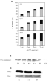
(A) Apoptosis and necrosis rates in 4HPR-treated CCRF–CEM cells. Cells were treated with increasing doses of 4HPR for 8, 12 and 24 h and then labelled with Annexin-V and Propidium Iodide as described in Materials and Methods. Apoptosis percentages are represented in grey, and in black percentages of necrotic cells in the same cultures. (B) Activation of caspase-3 in 4HPR-treated human CCRF–CEM cells. Blot shows the progressive pro-caspase-3 cleavage along time in 10 μM 4HPR-treated cells. (C) Bcl-2 and Bax protein expression in control and 4HPR-treated CEM cells.
Figure 4
Figure 4
Effect of antioxidants on the generation of reactive oxygen species in 4HPR-treated CCRF–CEM cells. A dose-dependent study. Cells were treated with or without increasing doses of 4HPR for 30 min and then ROS production was measured. In some cultures, the antioxidants NAC and vitamin E were added 2 h before initiating the treatments. The mean±s.d. of three experiments are shown. Differences in ROS production between antioxidant-treated cells and non-treated cells were statistically significant (P<0.01).
Figure 5
Figure 5
Effect of antioxidants on the survival of 4HPR-treated CEM cells. Cells were treated with 4HPR alone, or in combination with 10 mM NAC or 100 μM vitamin E. Relative percentages with respect to non-treated cells are shown. The means±s.d. of three experiments are shown. Differences in cell survival of antioxidant-treated cells were statistically significant with reference to those only treated with 4HPR (*P<0.05; **P<0.01).
Figure 7
Figure 7
Mitochondrial membrane potential in CCRF-cells treated with 4HPR. Cells treated with 6 μg ml−1 4HPR during short periods of time (1, 3 and 4 h) were labelled with Rhodamine 123 and analysed by flow cytometry. The reduction of transmembrane potential after 4 h of treatment is represented as a shift of the fluorescence peak to lower levels and the percentage of cells in the lower fluorescence category was plotted in the graphs.
Figure 8
Figure 8
Effect of vitamin E on 4HPR-induced cytochrome c release in human CCRF–CEM cells. Cells were incubated without (control) or with 5 μM 4HPR during 2, 8 and 12 h. In some cultures vitamin E was added 2 h before addition of 4HPR.

Similar articles

Cited by

References

    1. DeliaDAielloALombardiLPelicciGGrignaniFGrignaniFFormelliFMenardSCostaSCostaAVeronesiUPierottiMA1993N-(4-Hydroxyphenyl)retinamide induces apoptosis of malignant hemopoietic cell lines including those unresponsive to retinoic acid Cancer Res 5360366041 - PubMed
    1. DeliaDAielloAMeroniLNicoliniMReedJCPierottiMA1997Role of antioxidants and intracellular free radicals in retinamide-induced cell death Carcinogenesis 18943948 - PubMed
    1. Fernández-ChecaJCGarcía-RuizCColellAMoralesAMaríMMirandaMArditeE1998Oxidative stress: role of mitochondria and protection by glutathione BioFactors 8711 - PubMed
    1. FormelliFClericiMCampaTDi MauroMGMagniAMascottiG1993Five-year administration of fenretinide: pharmacokinetics and effects on plasma retinol concentrations J Clin Oncol 1120362042 - PubMed
    1. HailNLotanR2001Mitochondrial respiration is uniquely associated with the prooxidant and apoptotic effects of N-(4-hydroxyphenyl)retinamide J Biol Chem 2764561445621 - PubMed

Publication types

MeSH terms