

A novel combretastatin A-4 derivative, AC7700, strongly stanches tumour blood flow and inhibits growth of tumours developing in various tissues and organs
- PMID: 12085211
- PMCID: PMC2746587
- DOI: 10.1038/sj.bjc.6600296
A novel combretastatin A-4 derivative, AC7700, strongly stanches tumour blood flow and inhibits growth of tumours developing in various tissues and organs
Abstract
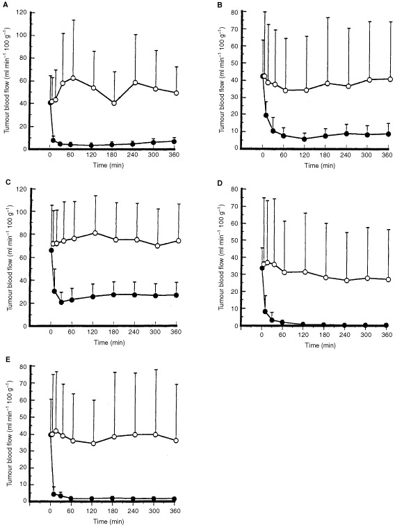
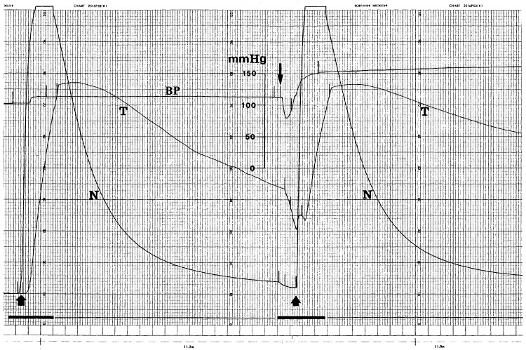
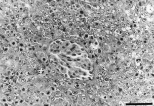
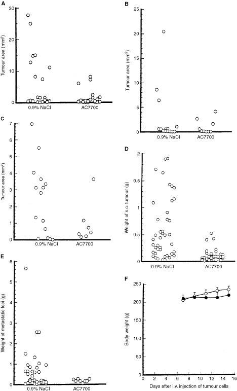
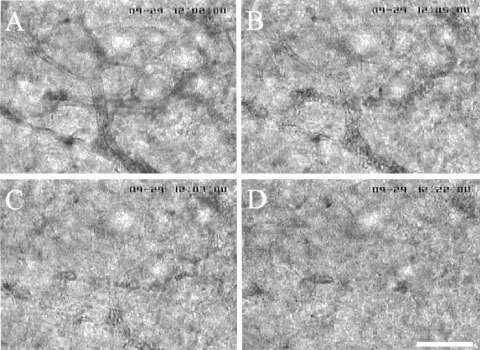
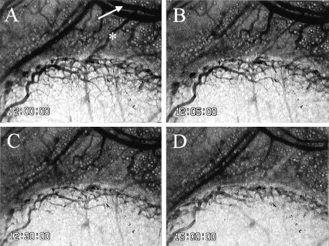
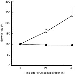
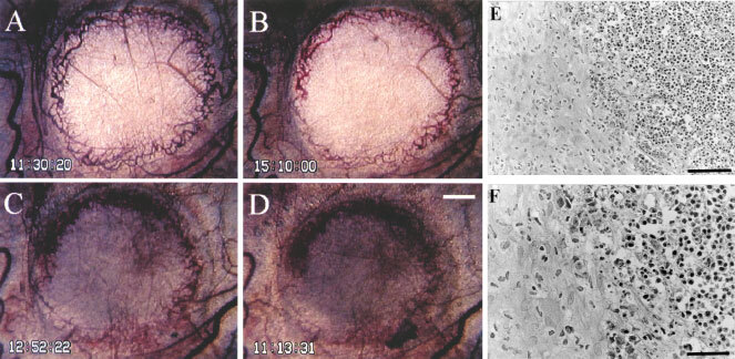
In a previous study, we used subcutaneous LY80 tumours (a subline of Yoshida sarcoma), Sato lung carcinoma, and methylcholanthrene-induced primary tumours, to demonstrate that a novel water-soluble combretastatin A-4 derivative, AC7700, abruptly and irreversibly stopped tumour blood flow. As a result of this interrupted supply of nutrients, extensive necrosis was induced within the tumour. In the present study, we investigated whether AC7700 acts in the same way against solid tumours growing in the liver, stomach, kidney, muscle, and lymph nodes. Tumour blood flow and the change in tumour blood flow induced by AC7700 were measured by the hydrogen clearance method. In a model of cancer chemotherapy against metastases, LY80 cells (2x10(6)) were injected into the lateral tail vein, and AC7700 at 10 mg x kg(-1) was injected i.v. five times at intervals of 2 days, starting on day 7 after tumour cell injection. The number and size of tumours were compared with those in the control group. The change in tumour blood flow and the therapeutic effect of AC7700 on microtumours were observed directly by using Sato lung carcinoma implanted in a rat transparent chamber. AC7700 caused a marked decrease in the tumour blood flow of all LY80 tumours developing in various tissues and organs and growth of all tumours including lymph node metastases and microtumours was inhibited. In every tumour, tumour blood flow began to decrease immediately after AC7700 administration and reached a minimum at approximately 30 min after injection. In many tumour capillaries, blood flow completely stopped within 3 min after AC7700 administration. These results demonstrate that AC7700 is effective for tumours growing in various tissues and organs and for metastases. We conclude that tumour blood flow stanching induced by AC7700 may become an effective therapeutic strategy for all cancers, including refractory cancers because the therapeutic effect is independent of tumour site and specific type of cancer.
comCopyright 2002 Cancer Research UK
Figures
Similar articles
-
Microvascular mechanisms by which the combretastatin A-4 derivative AC7700 (AVE8062) induces tumour blood flow stasis.Br J Cancer. 2003 Oct 6;89(7):1334-44. doi: 10.1038/sj.bjc.6601261. Br J Cancer. 2003. PMID: 14520469 Free PMC article.
-
Antitumor effects due to irreversible stoppage of tumor tissue blood flow: evaluation of a novel combretastatin A-4 derivative, AC7700.Jpn J Cancer Res. 1999 Sep;90(9):1026-38. doi: 10.1111/j.1349-7006.1999.tb00851.x. Jpn J Cancer Res. 1999. PMID: 10551334 Free PMC article.
-
Stoppage of blood flow in 3-methylcholanthrene-induced autochthonous primary tumor due to a novel combretastatin A-4 derivative, AC7700, and its antitumor effect.Med Sci Monit. 2001 Jan-Feb;7(1):26-33. Med Sci Monit. 2001. PMID: 11208488
-
Antineoplastic strategy: irreversible tumor blood flow stasis induced by the combretastatin A-4 derivative AVE8062 (AC7700).Chemotherapy. 2005 Oct;51(6):357-60. doi: 10.1159/000088961. Epub 2005 Oct 14. Chemotherapy. 2005. PMID: 16227690 Review.
-
[Tumor microcirculation and selective enhancement of drug delivery--clinical applications based on pathophysiological experiments].Gan To Kagaku Ryoho. 2000 Jul;27(8):1191-200. Gan To Kagaku Ryoho. 2000. PMID: 10945016 Review. Japanese.
Cited by
-
KPU-300, a Novel Benzophenone-Diketopiperazine-Type Anti-Microtubule Agent with a 2-Pyridyl Structure, Is a Potent Radiosensitizer That Synchronizes the Cell Cycle in Early M Phase.PLoS One. 2015 Dec 30;10(12):e0145995. doi: 10.1371/journal.pone.0145995. eCollection 2015. PLoS One. 2015. PMID: 26716455 Free PMC article.
-
Microtubule depolymerizing vascular disrupting agents: novel therapeutic agents for oncology and other pathologies.Int J Exp Pathol. 2009 Jun;90(3):284-94. doi: 10.1111/j.1365-2613.2009.00651.x. Int J Exp Pathol. 2009. PMID: 19563611 Free PMC article. Review.
-
Strategies for the Optimization of Natural Leads to Anticancer Drugs or Drug Candidates.Med Res Rev. 2016 Jan;36(1):32-91. doi: 10.1002/med.21377. Epub 2015 Sep 11. Med Res Rev. 2016. PMID: 26359649 Free PMC article. Review.
-
Induction of tumour blood flow stasis and necrosis: a new function for epinephrine similar to that of combretastatin A-4 derivative AVE8062 (AC7700).Br J Cancer. 2004 Jan 26;90(2):549-53. doi: 10.1038/sj.bjc.6601582. Br J Cancer. 2004. PMID: 14735207 Free PMC article.
-
Microvascular mechanisms by which the combretastatin A-4 derivative AC7700 (AVE8062) induces tumour blood flow stasis.Br J Cancer. 2003 Oct 6;89(7):1334-44. doi: 10.1038/sj.bjc.6601261. Br J Cancer. 2003. PMID: 14520469 Free PMC article.
References
-
- AlgireGHLegallaisFYAndersonBF1954Vascular reaction of normal and malignant tissues in vivo. VI. The role of hypotension in the action of components of podophyllin on transplantable sarcomas J Natl Cancer Inst 14879893 - PubMed
-
- AuklandKBowerBFBerlinerRW1964Measurement of local blood flow with hydrogen gas Circ Res 14164187 - PubMed
-
- BaguleyBCHoldawayKMThomsenLLZhuangLZwiLJ1991Inhibition of growth of colon 38 adenocarcinoma by vinblastine and colchicine: evidence for a vascular mechanism Eur J Cancer 27482487 - PubMed
-
- BibbyMCDoubleJALoadmanPMDukeCV1989Reduction of tumor blood flow by flavone acetic acid: a possible component of therapy J Natl Cancer Inst 81216220 - PubMed
Publication types
MeSH terms
Substances
LinkOut - more resources
Full Text Sources
Other Literature Sources
Medical
Research Materials
