. 2002 Jul;9(14):922-31.
doi: 10.1038/sj.gt.3301714.

Apical barriers to airway epithelial cell gene transfer with amphotropic retroviral vectors

Affiliations

Apical barriers to airway epithelial cell gene transfer with amphotropic retroviral vectors

G Wang et al. Gene Ther. 2002 Jul.

Abstract

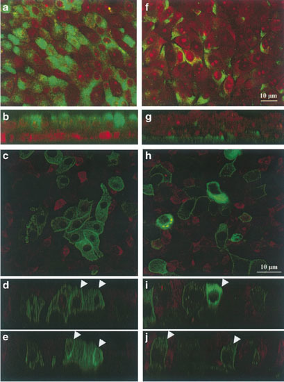
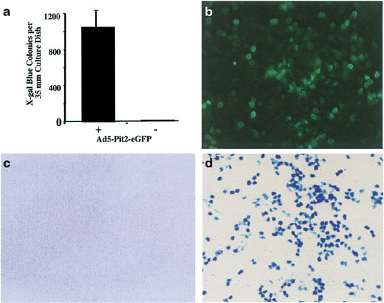
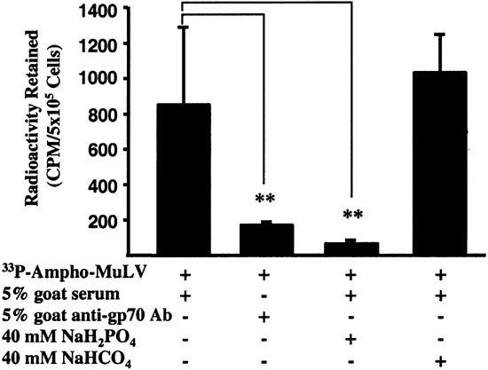
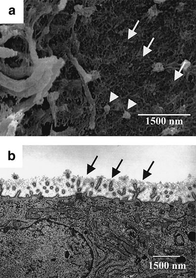
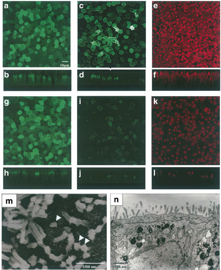
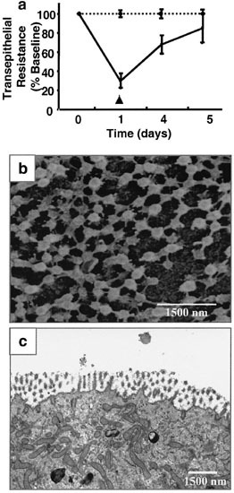
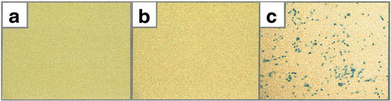
Gene transfer to airway epithelia with amphotropic pseudotyped retroviral vectors is inefficient following apical vector application. To better understand this inefficiency, we localized the expression of Pit2, the amphotropic receptor, in polarized human airway epithelia. Pit2 was expressed on both the apical and basolateral surfaces of the cells, suggesting that factors other than receptor abundance may limit apical gene transfer efficiency. Binding studies performed with radiolabeled amphotropic MuLV suggested that the apically applied virus binds to Pit2. Hypothetical barriers to retroviral gene transfer include the apical glycocalyx and other secreted products of epithelia. In this study, we demonstrated that sialic acid, keratan sulfate and collagen type V are present on the apical surface of well-differentiated human airway epithelia. While enzyme treatment reduced the abundance of these components, the treatment also decreased the transepithelial resistance to approximately 35% of the controls, suggesting that the epithelial integrity was impaired. To attain an airway epithelial culture with a modified apical surface and intact epithelial integrity, we utilized 100 mM 2-deoxy-D-glucose, a glycosylation inhibitor, to prevent the glycocalyx from reforming following enzyme treatment. This approach allowed the resistance, but not the apical glycocalyx to recover. Despite this physical modification of the cell surface, the amphotropic retroviral vector failed to transduce airway epithelia following apical application. These results suggest that factors other than apical receptor abundance and the glycocalyx inhibit amphotropic retroviral gene transfer in human airway epithelia.


Figures

Figure 1
Figure 1
Localization of Pit2 receptor expression on the apical and basolateral membranes of differentiated human airway epithelia. (a, b, f, g) Confocal micrographs of Pit2 immunolocalization on the apical and basolateral surfaces of polarized epithelia. (a, b) Apical application of Pit2 antibody. (f, g) Basolateral application of Pit2 antisera. (a and f) X-Y sections. (b and g) X-Z sections. Pit2 expression (FITC signal, green) is detected on both the apical and basolateral surfaces. Propidium iodide (red signal) counterstains nuclei and cytoplasm. (c–e and h–j) Confocal micrographs of airway epithelia expressing a Pit2-eGFP fusion receptor protein. Airway epithelia were infected with 50 MOI of Ad5CMVPit2-eGFP vector from the basolateral surface. Forty-eight hours later cells were examined for GFP localization by confocal microscopy (c–e, view 1; h–j, view 2; c and h, X-Y sections; d, e, i and j, X-Z sections). Pit2-eGFP expression (green signal) is seen on both the apical and basolateral membranes, as well as in the cytoplasm. Arrowheads indicate apical Pit2 signal. Views shown are representative of two different epithelial cell preparations examined.
Figure 2
Figure 2
Ad5-CMVPit2-eGFP vector transduction confers expression of functional GPF tagged Pit2. CHO cells were pre-infected with 50 MOI of Ad5-CMVPit2-eGFP 24 h before ampho MuLV infection. In negative controls, Ad5-CMVPit2-eGFP was omitted. Three days after the ampho MuLV infection, X-gal staining was performed to evaluate the transduction efficiency. As shown in panel (a), the pre-infection with Ad5-CMVPit2-eGFP reversed the insusceptibility of CHO cells to ampho-MuLV, demonstrating that the Pit2-eGFP fusion construct is functional. Similarly, we infected KGF-stimulated human airway cells with 50 MOI of Ad5-CMVPit2-eGFP. As seen in panel (b), a large population of airway cells expresses the Pit2-eGFP protein. However, when 20 MOI of ampho MuLV was applied to the apical surface, no gene transfer was noted after X-gal staining (c). If the MuLV vector was formulated with EGTA to disrupt tight junctions, efficient gene transfer was achieved (d).
Figure 3
Figure 3
Amphotropic MuLV vector binds specifically to the apical Pit2 receptor. The amphotropic MuLV vector was labeled with 33P-uridine as described in Materials and methods and applied to the apical surface under several conditions. As shown in the left-most bar in the figure, the radiolabeled vector binds to the apical surface. This binding was blocked by goat anti-gp70 antiserum (second bar), but not normal goat serum (first bar). Furthermore, 40 mM NaH2PO4 inhibited binding of amphotropic MuLV binding (third bar). In contrast, 40 mM NaHCO4 had no inhibitory effect (fourth bar). **, Indicates statistically significant difference compared with amphotropic MuLV + normal goat serum condition (P < 0.05, t test). Representative figure of four similar experiments.
Figure 4
Figure 4
Morphology of the apical surface of well-differentiated human airway epithelia. (a) SEM view of the apical surface. A dense fibrous network was noted around cilia and microvilli on the apical surface (arrows). Spherical bodies suggestive of glycocalyceal bodies were also seen (arrow heads). (b) Ultra-thin sections of the airway epithelia were examined by transmission electron microscopy. The fuzzy apical glycocalyx around and in between the microvilli is indicated by the arrows. Views shown are representative of three different epithelial cell preparations examined.
Figure 5
Figure 5
Components of the apical glycocalyx and surface alterations produced by enzyme treatment. Confocal micrographs (a–l). Apical surface staining for sialic acids in control airway cells (a, b) and enzyme-treated airway cells (g, h) with WGA-FITC as described in Materials and methods. Apical keratan sulfate staining of the control airway epithelia (c, d) and enzyme-treated cells (i, j). Apical localization of collagen type V in control (e, f) and enzyme-treated airway epithelia (k, l). X-Y sections (a, c, e, g, i and k); X-Z sections (b, d, f, h, j and l); (m–n) Airway epithelia treated with enzymes were examined by scanning and transmission electron microscopy; m, scanning EM image shows reduction in the fibrous network of the apical glycocalyx. Some spherical bodies suggestive of glycocalyceal bodies remain (arrow heads). n, Transmission EM image demonstrates that the fuzzy material around microvilli and cilia was much reduced. Contrast panels (m) and (n) with images in Figure 4. Views shown are representative of three different epithelial cell preparations examined.
Figure 6
Figure 6
Enzyme treatment significantly decreases the transepithelial resistance. Following enzyme treatment for 2 h at 37°C, the transepithelial resistance was measured as described in Materials and methods. As compared with controls, this treatment reduced the transepithelial resistance to ~35% of the controls. The transepithelial resistance of enzyme-treated cells gradually recovered to baseline values after 4–5 days (data not shown). Results shown represent mean ± s.e. *, Indicates statistically significant difference compared with controls (P < 0.05, t test, n = 4).
Figure 7
Figure 7
Glycosylation inhibition following enzyme treatment allows transepithelial resistance, but not the apical glycocalyx, to recover. 100 mM 2-deoxy-D-glucose was applied to the culture medium after the apical surface of differentiated airway epithelia was digested enzymatically. Transepithelial resistance was measured each day for 5 days. (a) In enzyme-treated cells transepithelial resistance gradually recovered to control levels 5 days after treatment. Results shown represent mean ±s.e. (b) Scanning electron microscopy demonstrates that the apical surface remained modified after enzyme treatment and culture in 2-deoxy-D-glucose-containing media. The fibrous network of the apical glycocalyx remains reduced. Some spherical bodies suggestive of glycocalyceal bodies remain. (c) Transmission EM similarly shows that the glycocalyx is largely under these conditions. Views shown are representative of three different epithelial cell preparations examined.
Figure 8
Figure 8
Modification of the apical glycocalyx fails to facilitate gene transfer from the apical surface with amphotropic MuLV vector. All cells were pretreated with KGF to stimulate proliferation. Views shown are low, power en face photomicrographs of X-gal stained epithelia. (a) 20 MOI of amphotropic MuLV was applied apically to control airway epithelia and no significant gene transfer occurred. (b) Enzyme and 2-deoxy-D-glucose-treated airway epithelia were transduced apically with 20 MOI of the amphotropic retroviral vector. There was no evidence of gene transfer. (c) Enzyme and 2-deoxy-D-glucose-treated airway epithelia were transduced apically with 20 MOI of the amphotropic MuLV vector formulated with 6 mM EGTA. X-gal positive (blue) cells representing gene transfer were only noted in cells treated with EGTA formulated vector. The experiment was performed three times with two different epithelial cell preparations.

Similar articles

Cited by

References

    1. Johnson LG. Efficiency of gene transfer for restoration of normal airway epithelial function in cystic fibrosis. Nature Genet. 1992;2:21–25. doi: 10.1038/ng0992-21. - DOI - PubMed
    1. Wang G, Sinn PL, McCray PB., Jr Development of retroviral vectors for gene transfer to airway epithelia. Curr Opin Mol Ther. 2000;2:497–506. - PubMed
    1. Flotte TR. Stable in vivo expression of the cystic fibrosis transmembrane conductance regulator with an adeno-associated virus vector. Proc Natl Acad Sci USA. 1993;90:10613–10617. doi: 10.1073/pnas.90.22.10613. - DOI - PMC - PubMed
    1. Pickles RJ. Retargeting the coxsackievirus and adenovirus receptor to the apical surface of polarized epithelial cells reveals the glycocalyx as a barrier to adenovirus-mediated gene transfer. J Virol. 2000;74:6050–6057. doi: 10.1128/JVI.74.13.6050-6057.2000. - DOI - PMC - PubMed
    1. Walters RW. Basolateral localization of fiber receptors limits adenovirus infection of airway epithelia. J Biol Chem. 1999;274:10219–10226. doi: 10.1074/jbc.274.15.10219. - DOI - PubMed

Publication types

MeSH terms