. 2002 Jun 18:3:18.
doi: 10.1186/1471-2091-3-18.

Insertion of a small peptide of six amino acids into the beta7-beta8 loop of the p51 subunit of HIV-1 reverse transcriptase perturbs the heterodimer and affects its activities

Affiliations

Insertion of a small peptide of six amino acids into the beta7-beta8 loop of the p51 subunit of HIV-1 reverse transcriptase perturbs the heterodimer and affects its activities

Pradeep K Pandey et al. BMC Biochem. .

Abstract

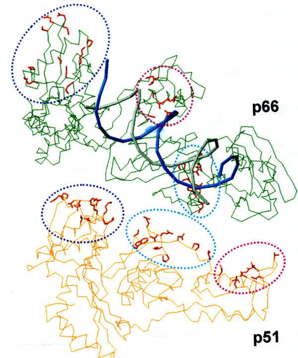
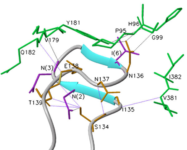
Background: HIV-1 RT is a heterodimeric enzyme, comprising of the p66 and p51 subunits. Earlier, we have shown that the beta7-beta8 loop of p51 is a key structural element for RT dimerization (Pandey et al., Biochemistry 40: 9505, 2001). Deletion or alanine substitution of four amino acid residues of this loop in the p51 subunit severely impaired DNA binding and catalytic activities of the enzyme. To further examine the role of this loop in HIV-1 RT, we have increased its size such that the six amino acids loop sequences are repeated in tandem and examined its impact on the dimerization process and catalytic function of the enzyme.

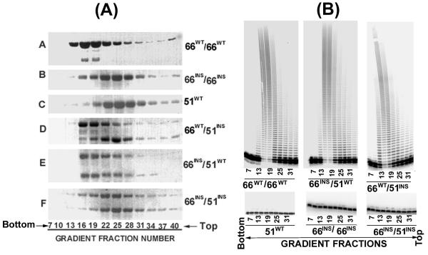
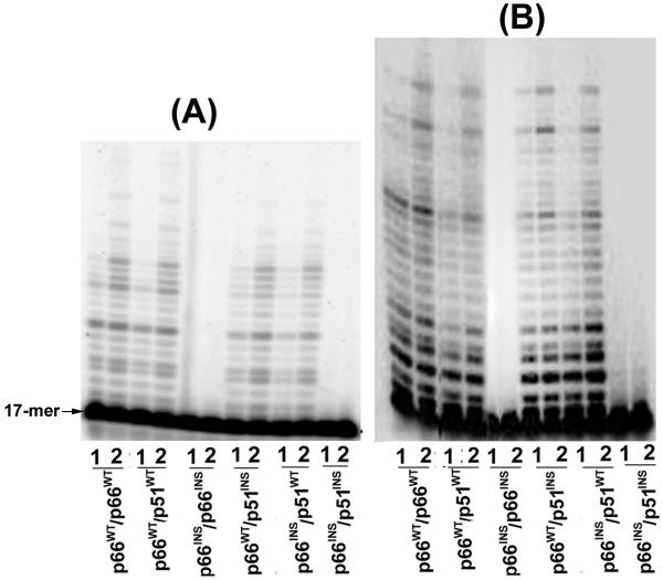
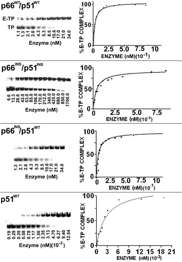
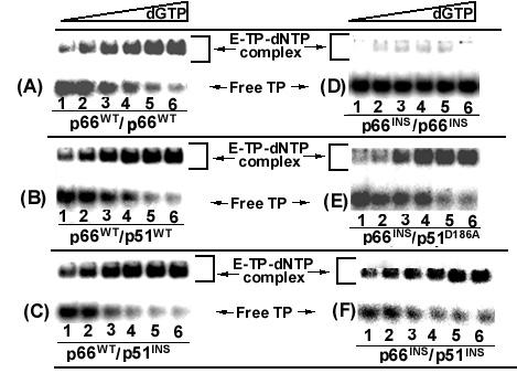
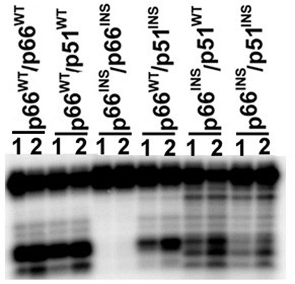
Results: The polymerase and the RNase H activities of HIV-1 RT carrying insertion in the beta7-beta8 loop of both the subunits (p66INS/p51INS) were severely impaired with substantial loss of DNA binding ability. These enzymatic activities were restored when the mutant p66INS subunit was dimerized with the wild type p51. Glycerol gradient sedimentation analysis revealed that the mutant p51INS subunit was unable to form stable dimer either with the wild type p66 or mutant p66INS. Furthermore, the p66INS/p66INS mutant sedimented as a monomeric species, suggesting its inability to form stable homodimer.

Conclusion: The data presented herein indicates that any perturbation in the beta7-beta8 loop of the p51 subunit of HIV-1 RT affects the dimerization process resulting in substantial loss of DNA binding ability and catalytic function of the enzyme.


Figures

Figure 1
Figure 1
(A) Sedimentation profile of the wild type and mutant HIV-1 RT. Mutant enzymes carrying 6 amino acids insertion in the β7-β8 loop of either or both the subunits were applied on 10–30% linear glycerol gradient (5 mL) and centrifuged at 48,000 rpm in SW 50.1 rotor for 22 hours. Gradients were fractionated from the bottom and subjected to SDS polyacrylamide gel electrophoresis and Coomassie Blue staining. (B) Polymerase Activity profile of the glycerol gradient fractions. Every third fraction between 7 and 33 of the glycerol gradient (Fig. 1A) was diluted 10-fold and analyzed for its polymerase activity on poly (rA)(dT)18 as described under Materials and Methods. The reaction products were resolved on an eight percent denaturing poly-acrylamide urea gel and subjected to PhosphorImaging.
Figure 2
Figure 2
Analysis of polymerase products catalyzed by insertion mutants of HIV-1 RT. Primer extension reactions catalyzed by the wild type and mutant enzymes were on DNA (panel A) and RNA (panel B) templates, primed with 5'-32P labeled 17-mer primer. Each set of reactions was carried out for 30 seconds (lane 1) and 60 seconds (lane 2) at 37°C and quenched by the addition of equal volume of Sanger's gel loading dye. The reaction products were resolved on an 8% polyacrylamide-7M urea gel and subjected to PhosphorImager analysis.
Figure 3
Figure 3
DNA binding affinity of mutant HIV-1 RT carrying insertion in the β7–β8 loop of either or both the subunits. The 49-mer DNA primed with 5'-32P labeled 21-mer DNA primer was incubated with varying concentrations of the individual enzyme at 4°C for 10 min. The mixture was electrophoresed under non-denaturing conditions on a 6% (w/v) polyacrylamide gel and analyzed on phosphorImager (Left panel). The positions of the free template primer (TP) and enzyme bound template-primer (E-TP binary complex) are indicated for the wild type enzyme. The percent of E-TP complex formed as a function of enzyme concentration was plotted for determining the Kd values (Right panel).
Figure 4
Figure 4
Apparent dNTP binding affinity of mutant HIV-1 RT carrying insertion in the β7–β8 loop of either or both the subunits. The 33-mer DNA primed with 5'-32P labeled dideoxy (ddC) terminated 21-mer DNA primer was incubated with the individual enzyme at 4°C for 10 min. The binding affinity [Kd (dNTP)] of the wild type enzyme and its mutant derivatives in the ternary complex was determined by incubating the preformed E-TP binary complex of the individual enzyme species at different concentrations (0.26–800 μM) of the next correct dNTP (dGTP). Following incubation with dGTP, 300 fold molar excess of the unlabeled TP was used as trap to remove the readily dissociable binary complexes from the ternary complex population. The extent of stable ternary complexes formed were resolved on 6% native polyacrylamide gel and analyzed by phosphorImaging. The [Kd [dNTP]], in the ternary complex for each enzyme was determined by quantifying the E-TP-dNTP ternary complexes as a function of increasing concentrations of dGTP and fitting the data to the equation for single-site ligand binding using Sigma-Plot. Lanes 1–6 represent the ternary complex formed in the presence of dGTP at 0.2, 1.28, 6.4, 32, 160 and 800 μM, concentrations.
Figure 5
Figure 5
RNase H activity of the wild-type HIV-1 RT and its mutant derivatives. The individual enzymes were incubated with 5'-32P-labeled 30-mer RNA annealed with 30-mer complementary DNA strand, at 37°C for 30 sec and 1 min as described in Materials and Methods. The cleavage products were resolved on an 8% denaturing polyacrylamide-urea gel and analyzed on a phosphorImager. Lanes 1 and 2 represent reactions carried out for 30 sec and 1 min, respectively.
Figure 6
Figure 6
Amino acid residues in the p51 subunit in contact with the p66 subunit. The Cα backbone of p51 and p66 is shown in orange and green, respectively. Amino acids residues of p51 making contact with the p66 subunit are represented as 'sticks' and are shown encircled. The location of analogous residues in p66 are also encircled. It may be noted that these residues in p66 are scattered. The residues circled within 'blue' are from fingers; those in light blue' are from 'connection' and those in pink are from 'thumb' subdomains. The template (blue) and primer (gray) are also displayed in this figure. The residues in blue circle are V21, K22, P25, P52, E53, N57, T131, N136, N137, E138, T139, in light blue circle are P392, I393, Q394, E396, T397, T400, W401, N418, P420, L422, and those in pink circle are N255, Q258, V261, N265, V276, L283, T286, L289.
Figure 7
Figure 7
Molecular model of the inserted peptide in the β7–β8 loop of the p51 subunit. Molecular modeling of the extended loop was performed by the 'Loop Search' option of SYBYL version 6.5 (Tripos Associates, St. Louis, MO). A total of 100 loops, matching the Cα distances constrain between the end points of the loop residues, were obtained. The loops were examined for 'bumps' with the neighboring protein structure. Selection of the appropriate loop was based on the least root mean square deviation and maximum homology with the inserted loop sequence. A loop satisfying these criteria containing two small anti-parallel β-strands is shown in cyan. The side chains of amino acid residues of p66 are shown in green and those of p51 (wild type) are shown in orange. The side chains of interacting residues in the inserted peptide are colored violet and are indicated by their position number in parentheses. Interactions seen in the wild type loop are shown in blue dotted line while additional interactions are shown in black dotted lines.

Similar articles

Cited by

References

    1. Chandra A, Gerber T, Chandra P. Biochemical heterogeneity of reverse transcriptase purified from the AIDS virus, HTLV-III, FEBS Lett. 1986;197:84–88. doi: 10.1016/0014-5793(86)80303-9. - DOI - PubMed
    1. Muller B, Restle T, Weiss S, Gautel M, Sczakiel G, Goody R. Co-expression of the subunits of the heterodimer of HIV-1 reverse transcriptase in Escherichia coli. J Biol Chem. 1989;264:13975–13978. - PubMed
    1. Baltimore D. RNA-dependent DNA polymerase in virions of RNA tumor Viruses. Nature. 1970;226:1209–1211. - PubMed
    1. Temin HM, Mizytani S. RNA-directed DNA polymerase in virions of Rouse sarcoma virus. Nature. 1970;226:2111–1213. - PubMed
    1. Farmerie WG, Leob D, Casavant NC, Hutchinson CAd, Egell MH, Swanstrom R. Expression and processing of AIDS virus reverse transcriptase in E. coli. Science. 1987;236:305–308. - PubMed

Publication types

MeSH terms

LinkOut - more resources