. 2002 Jun 19:3:7.
doi: 10.1186/1471-2202-3-7.

A new way to rapidly create functional, fluorescent fusion proteins: random insertion of GFP with an in vitro transposition reaction

Affiliations

A new way to rapidly create functional, fluorescent fusion proteins: random insertion of GFP with an in vitro transposition reaction

Douglas L Sheridan et al. BMC Neurosci. .

Abstract

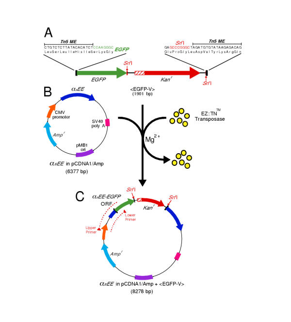
Background: The jellyfish green fluorescent protein (GFP) can be inserted into the middle of another protein to produce a functional, fluorescent fusion protein. Finding permissive sites for insertion, however, can be difficult. Here we describe a transposon-based approach for rapidly creating libraries of GFP fusion proteins.

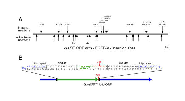
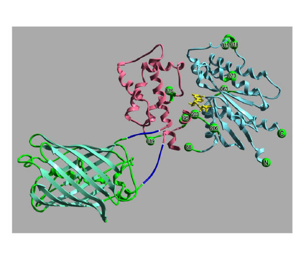
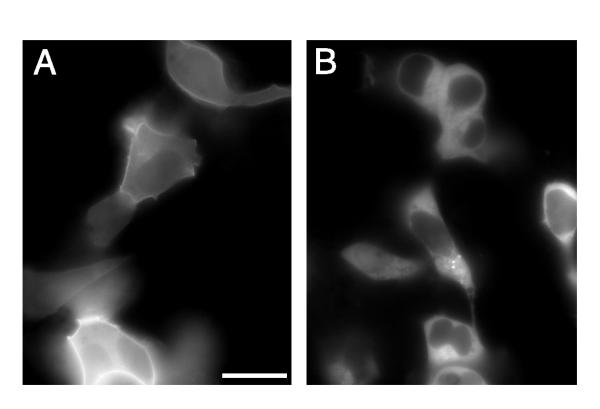
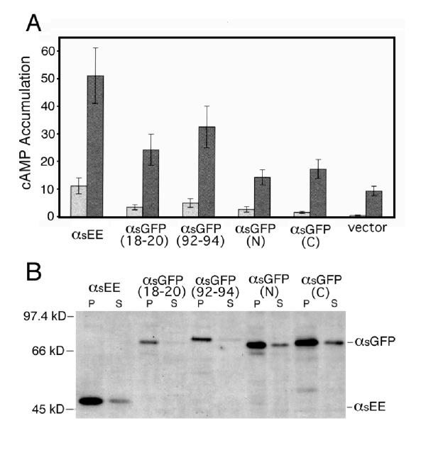
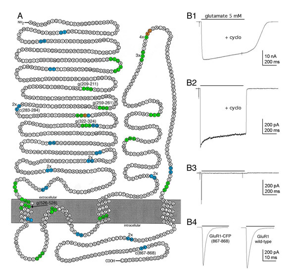
Results: We tested our approach on the glutamate receptor subunit, GluR1, and the G protein subunit, alphas. All of the in-frame GFP insertions produced a fluorescent protein, consistent with the idea that GFP will fold and form a fluorophore when inserted into virtually any domain of another protein. Some of the proteins retained their signaling function, and the random nature of the transposition process revealed permissive sites for insertion that would not have been predicted on the basis of structural or functional models of how that protein works.

Conclusion: This technique should greatly speed the discovery of functional fusion proteins, genetically encodable sensors, and optimized fluorescence resonance energy transfer pairs.


Figures

Figure 1
Figure 1
Transposition with <EGFP-V> (A) The transposon, <EGFP-V>, is flanked by 19 bp inverted repeats, the MEs. The EGFP coding region is positioned such that when <EGFP-V> inserts between the codons of a target gene, a fusion protein will be produced. <EGFP-V> also carriesKanr. There is a stop codon in the 5' end of the Kanr cassette in the same frame as the EGFP coding sequence, so if the transposon lands in an open reading frame, in the correct orientation and frame, a truncated, EGFP-tagged, protein will initially be produced. Removal of theKanr cassette by Srf I digestion and re-ligation produces a reading frame that extends across the entire transposon. (B) The target plasmid, αsEE in pcDNA1/Amp, encodes an epitope tagged version of the G protein subunit αs. (C) Transposed plasmids carry Ampr and Kanr. <EGFP-V> insertions within the target gene produce a PCR product when <EGFP-V> is inserted in the correct orientation, and the size of the PCR product reveals which <EGFP-V> insertions are in the coding sequence.
Figure 2
Figure 2
<EGFP-V> Insertions in αsEE. (A) Location of 35 <EGFP-V> insertions in the αsEE coding region. The 13 labeled insertions marked above the coding region are in the correct reading frame and encode fluorescent fusion proteins (12 unique insertions, 1 duplication). The insertions are named for the 3 amino acids of αs duplicated during transposition. The 22 unlabeled insertions (18 unique) marked below the coding region are those in which <EGFP-V> landed out-of-frame with respect to αsEE. Redundant insertions are indicated by the number of clones recovered at that site (e.g. 2X, 4X). (B) Srf I digestion of the transposed clone, followed by religation, removes theKanr selection cassette and produces the full-length fusion protein. In the final fusion protein, the EGFP domain is bordered by amino acid linkers encoded by the Tn5 MEs as well as the 9 bp duplication of the target sequence that is generated during the transposition process.
Figure 3
Figure 3
Model of αs-GFP(92–94). The GFP insertions into αs can be interpreted in the context of the structures of GFP (PDB file: 1EMA) and αs-GTPγ S (PDB file: 1AZT). In this image, the structure of GFP [42] is green, while the helical domain of the αsubunit [15] is pink, and the GTPase domain is blue. GTPγ S is yellow. The GFP insertion αs-GFP(92–94) that produced a functional G protein subunit is illustrated by the short linkers (encoded by the Tn5 MEs) between GFP and αs (dark blue). The other sites of <EGFP-V> insertion are shown as green spheres. The numbers on the spheres indicate the second of the three duplicated residues that flank the transposon insertions (the numbers are based on the long form of αs).
Figure 4
Figure 4
Localization of αs-GFP Fusion Proteins in Living Cells. (A) Membrane localization of tribrid fusion, αs-GFP(92–94), in HEK 293 cells ~24 hr after co-transfection with β1 and γ7. Similar localization patterns were observed when αs-GFP(18–20), αs-GFP(N), or αs-GFP(C) fusions were co-expressed with β1 and γ7 (A non-linear representation of the image brightness was used to illustrate both the dimly fluorescent cells in the upper right corner and the very bright ones at the bottom, scale bar = 20 μm). (B) The remaining 10 tribrid fusion proteins were evenly distributed throughout the cytosol (with little fluorescence in the nucleus) as seen here in HEK 293 cells transiently expressing αs-GFP(362–364), β1 and γ7.
Figure 5
Figure 5
Activity and Expression Levels of αs-GFP Fusion Proteins. (A) HEK 293 cells were transfected with 2 μg/106 cells of the indicated αs-GFP constructs or vector alone (pcDNA1/Amp) and 0.2 μg/106 cells of plasmid encoding the LH receptor. cAMP accumulation was measured in the presence (dark gray bars) or absence (light gray bars) of hCG, an LH receptor agonist. Cells expressing each of the αs-GFP fusion proteins exhibited increased basal and stimulated cAMP accumulation relative to cells expressing vector alone, but only the increases in cells expressing αs-GFP(92–94) were significantly greater (p < .05). Values represent the mean ± S.E. of 5 independent experiments. (B) Immunoblots of the membrane pellets (P) and supernatant (S) fractions from transiently transfected HEK 293 cells. Expression levels of tribrid fusion proteins αs-GFP(18–20) and αs-GFP(92–94), but not of amino- and carboxy-terminus fusions, αs-GFP(N) and αs-GFP(C), respectively, were decreased in both fractions relative to that of unlabeled αsEE. Similar results were obtained in an additional experiment.
Figure 6
Figure 6
Insertion Sites and Functional Screening of GluR1-GFP Fusion Proteins (A) A model of GluR1 topology showing the locations of the GFP insertion in 45 fluorescent fusion proteins. In-frame insertions of <TgPT-0> result in a 3 amino acid duplication (green) flanking the insertion site. In-frame insertions of <TcPT-1> generate only a 2 amino acid duplication (cyan) in the target because of the different reading frame. The orange amino acids are overlapping insertion sites of the two transposons (See supplemental diagram for the two reading frames). Multiple clones with identical transpositions are identified as 2x, 3x, etc. The six insertions resulting in functional, fluorescent GluR1-GFP/CFP tribrid fusion proteins are identified by the duplicated target amino acids (e.g. g209–211, c867–868). This figure was adapted from [43]. (B) AMPA receptor-mediated current from GluR1-CFP(867–868). (B1) Large whole-cell current elicited by the rapid sustained application of 5 mM glutamate (bar) in a cell transiently expressing GluR1-CFP(867–868) after reducing desensitization with cyclothiazide (100 μM). (B2) Current elicited by 5 mM glutamate in an outside-out patch pulled from a cell transiently expressing GluR1-CFP(867–868). (B3) Current elicited in the same patch as B3, but in the absence of cyclothiazide. Note the rapid and nearly complete desensitization of the current. (B4) The trace on the left is an expanded time scale of B3, the trace on the right is from an outside-out patch pulled from a cell transiently expressing wild-type GluR1 in the presence of 5 mM glutamate without cyclothiazide.

Similar articles

Cited by

References

    1. Chalfie M, Tu Y, Euskirchen G, Ward WW, Prasher DC. Green fluorescent protein as a marker for gene expression. Science. 1994;263:802–5. - PubMed
    1. Wang S, Hazelrigg T. Implications for bcd mRNA localization from spatial distribution of exu protein in Drosophila oogenesis. Nature. 1994;369:400–3. doi: 10.1038/369400a0. - DOI - PubMed
    1. Marshall J, Molloy R, Moss GW, Howe JR, Hughes TE. The jellyfish green fluorescent protein: a new tool for studying ion channel expression and function. Neuron. 1995;14:211–5. - PubMed
    1. Chalfie M. Green fluorescent protein. Photochem Photobiol. 1995;62:651–6. - PubMed
    1. Siegel MS, Isacoff EY. A genetically encoded optical probe of membrane voltage. Neuron. 1997;19:735–41. - PubMed

Publication types

LinkOut - more resources