. 2002 Feb;1(1):53-62.
doi: 10.1016/s1535-6108(01)00002-2.

Induction of ovarian cancer by defined multiple genetic changes in a mouse model system

Affiliations

Induction of ovarian cancer by defined multiple genetic changes in a mouse model system

Sandra Orsulic et al. Cancer Cell. 2002 Feb.

Abstract

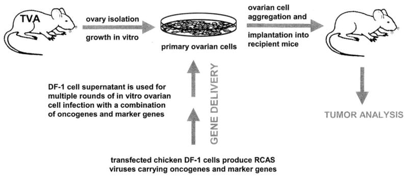
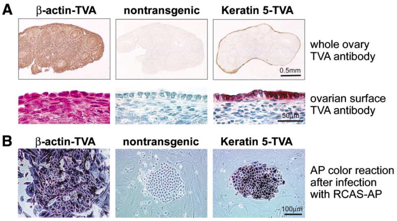
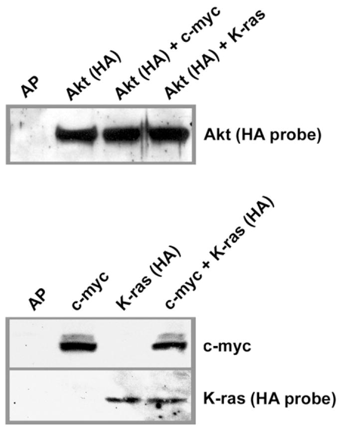
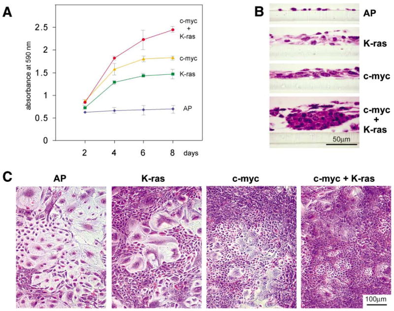
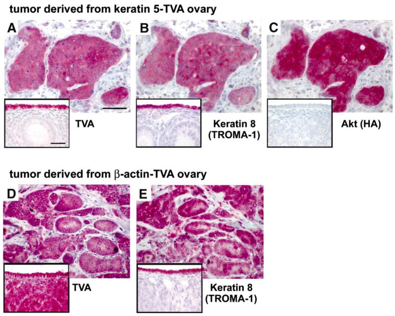
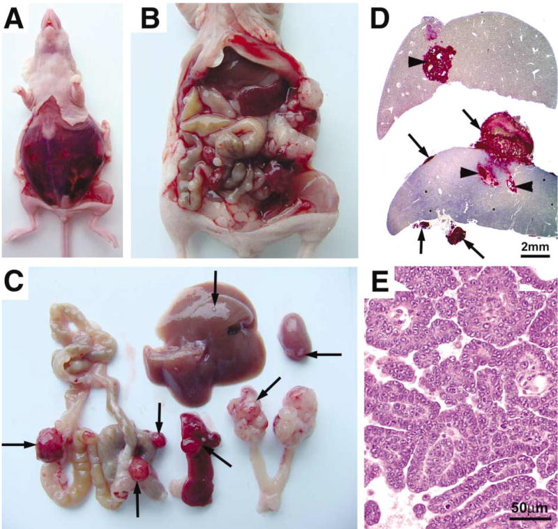
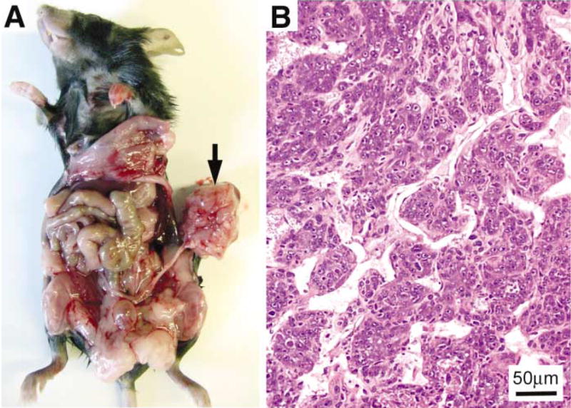
We have developed a mouse model for ovarian carcinoma by using an avian retroviral gene delivery technique for the introduction of multiple genes into somatic ovarian cells of adult mice. Ovarian cells from transgenic mice engineered to express the gene encoding the avian receptor TVA were efficiently infected in vitro with multiple vectors carrying coding sequences for oncogenes and marker genes. When target cells were derived from TVA transgenic mice deficient for p53, the addition of any two of the oncogenes c-myc, K-ras, and Akt were sufficient to induce ovarian tumor formation when infected cells were injected at subcutaneous, intraperitoneal, or ovarian sites. We demonstrated that the ovarian surface epithelium is the precursor tissue for these ovarian carcinomas, and that introduction of oncogenes causes phenotypic changes in the ovarian surface epithelial cells. The induced ovarian tumors in mice resembled human ovarian carcinomas in their rapid progression and intraperitoneal metastatic spread.


Figures

Figure 1
Figure 1
Strategy for viral gene delivery to ovarian cells in vitro and introduction of infected cells into recipient mice Ovaries are isolated from a TVA transgenic mouse to restrict infection to ovarian cells. DF-1 chicken cells transfected with an RCAS vector carrying a gene of interest are used to obtain high titer virus stock. In vitro propagation of primary ovarian cells facilitates multiple rounds of infection with a combination of RCAS viruses carrying different oncogenes and marker genes. After several days of infection in vitro, ovarian cells are aggregated and implanted into recipient mice.
Figure 2
Figure 2
Expression of TVA and susceptibility of ovarian cells from TVA transgenic mice to RCAS infection A: Immunohistochemical detection of the TVA receptor with the TVA antibody. Ovaries isolated from β-actin-TVA transgenic mice express the TVA receptor in both stromal cells and the ovarian surface epithelium. In ovaries isolated from keratin 5-TVA transgenic mice, expression of the TVA receptor is restricted to the cells of the ovarian surface epithelium. B: Detection of RCAS-AP-infected cells by AP color reaction. Ovarian cells from each transgenic line were infected with the RCAS-AP virus in culture. Endogenous AP was heat-inactivated, and the infected cells that produce AP were detected by the AP color reaction. Both stromal and epithelial cells from β-actin-TVA ovaries are susceptible to RCAS-AP infection. In ovaries from keratin 5-TVA mice, only the ovarian surface epithelial cells are susceptible to RCAS-AP infection, while stromal cells are AP-negative.
Figure 3
Figure 3
Expression of oncogenic proteins in ovarian cells after in vitro infection with RCAS viruses Western blot of β-actin-TVA/p53−/− ovarian cells infected with RCAS viruses carrying AP, c-myc, K-ras, or Akt, individually or in combinations of two RCAS viruses. The same amount of cell lysate was loaded into each lane. HA-tagged Akt and K-ras proteins were detected with the anti-HA probe. The expression of c-myc was determined with an antibody against human c-myc.
Figure 4
Figure 4
Oncogenic c-myc and K-ras collaborate in inducing proliferation of epithelial ovarian cells A: Proliferation curves of ovarian cells from β-actin-TVA/p53−/− mice that were infected with RCAS viruses carrying AP, c-myc, K-ras, or with a combination of RCAS viruses carrying c-myc and K-ras. Five days after infection with RCAS viruses, each culture of the virally infected cells was split into 12 separate cultures that were maintained for 8 days. The cell proliferation rates were determined by fixing triplicate cultures every 2 days, staining them with crystal violet dye, and measuring the light absorbance of the extracted dye at 590 nm. This procedure allowed us to count the proliferating cells, as well as the viable terminally differentiated cells that would normally be lost if the cells were continually passaged. Proliferation curves were plotted as a ratio of the light absorbance and the time. The error bars indicate the extreme values of light absorbance in three separate cultures. Proliferation of ovarian cells from β-actin-TVA/p53−/− mice was induced by oncogenic change in either c-myc or K-ras, but the effect was further amplified in ovarian cells with genetic changes in both genes. B: Morphological appearance of the cell populations described in A at day 6. Cultured ovarian cells infected with RCAS-AP acquire features of normal terminal differentiation and senescence. Introduction of oncogenic lesions induces marked changes to ovarian cell morphology and differentiation, which is especially pronounced in the ovarian epithelial cells. C: Representative cross-sections of ovarian cell populations in B.
Figure 5
Figure 5
Ovarian surface epithelium is the precursor tissue for ovarian carcinoma A–C: Sections of a subcutaneous tumor in a nude mouse derived from the keratin 5-TVA/p53−/− ovarian cells infected with a combination of RCAS-c-myc and RCAS-Akt viruses. The consecutive sections were probed with antibodies that recognize TVA, Keratin 8 and the HA-tagged Akt protein. Insets show a normal ovary from an unin-fected keratin 5-TVA mouse probed with the same antibodies. A: The TVA-positive cells in the tumor originate from the ovarian surface epithelium because this is the only cell type that expresses TVA in the ovaries from keratin 5-TVA transgenic mice (A, inset). B: The same TVA-positive cells in the tumor are also positive for the Keratin 8 marker, indicating that they maintain their epithelial characteristics and resemble the Keratin 8-positive cells of the ovarian surface epithelium (B, inset). C: The TVA-positive cells in the tumor also express virally introduced Akt oncogene. The HA-tagged Akt is not present in the uninfected ovary (C, inset). D and E: Consecutive sections of a subcutaneous tumor in a nude mouse derived from the β-actin-TVA/p53−/− ovarian cells infected with a combination of RCAS-c-myc and RCAS-K-ras viruses. The sections were probed with antibodies that recognize TVA receptor and Keratin 8. Insets show a normal ovary from uninfected β-actin-TVA mouse probed with the same antibodies as the tumor sections. D: The tumor consists of TVA-positive and TVA-negative cells. The TVA-positive cells originate from the ovary of the β-actin-TVA transgenic mouse (D, inset). Since all cells from β-actin-TVA ovaries express TVA, the TVA-negative stromal cells in the tumor must be recruited from the host tissue and not from ovarian stroma. E: The TVA-positive cells in the tumor are epithelial as determined by the expression of the Keratin 8 marker which is present exclusively in the epithelial cells in the ovary (E, inset). Each bar represents 50 μm.
Figure 6
Figure 6
Intraperitoneal tumor spread in nude mice after implantation of ovarian cells with induced genetic lesions under the ovarian bursa A–E: Tumors derived from β-actin-TVA/p53−/− ovarian cells that were infected with a combination of RCAS viruses carrying c-myc and K-ras oncogenes and implanted under the ovarian bursa of a nude mouse. A: Abdominal bloating and the presence of bloody ascites were the typical signs of the intraperitoneal tumor spread. B: Ovarian tumor spread in the peritoneal cavity. C: Ovarian tumor spread to the intraperitoneal and retroperitoneal organs. Arrows indicate ovarian tumor spread to the spleen, liver, intestine, kidney, and the contralateral ovary. D: Sections of the liver and the spleen probed with antibody against TVA to identify the ovarian tumor cell metastases. The tumor cells metastasize to the surfaces of organs (arrows) and deep into the tissue (arrowheads). E: Papillary organization of metastatic ovarian tumor resembles ovarian papillary carcinoma.
Figure 7
Figure 7
Ovarian tumor induction in immunocompetent mice A: Ovarian cells from the right ovary of the β-actin-TVA/p53−/− mouse were transduced with c-myc, K-ras, and Akt oncogenes and implanted under the ovarian bursa of the left ovary. The arrow points to the tumor that developed in the left ovary three months after intrabursal implantation of the RCAS-infected ovarian cells. B: H&E staining of the tumor section reveals an undifferentiated epithelial neoplasm, arranged in nests.

Comment in

  • Of mice and women.
    Foulkes WD. Foulkes WD. Cancer Cell. 2002 Feb;1(1):11-2. doi: 10.1016/s1535-6108(02)00021-1. Cancer Cell. 2002. PMID: 12086881

References

    1. Adams AT, Auersperg N. Transformation of cultured rat ovarian surface epithelial cells by Kirsten murine sarcoma virus. Cancer Res. 1981;41:2063–2072. - PubMed
    1. Auersperg N, Wong AS, Choi KC, Kang SK, Leung PC. Ovarian surface epithelium: biology, endocrinology, and pathology. Endocr Rev. 2001;22:255–288. - PubMed
    1. Aunoble B, Sanches R, Didier E, Bignon YJ. Major oncogenes and tumor suppressor genes involved in epithelial ovarian cancer (review) Int J Oncol. 2000;16:567–576. - PubMed
    1. Baker VV, Borst MP, Dixon D, Hatch KD, Shingleton HM, Miller D. c-myc amplification in ovarian cancer. Gynecol Oncol. 1990;38:340–342. - PubMed
    1. Bates P, Young JA, Varmus HE. A receptor for subgroup A Rous sarcoma virus is related to the low density lipoprotein receptor. Cell. 1993;74:1043–1051. - PubMed

Publication types

MeSH terms