Comparative Study
. 2002 Jul;136(5):645-58.
doi: 10.1038/sj.bjp.0704734.

Involvement of NO in the failure of neutrophil migration in sepsis induced by Staphylococcus aureus

Affiliations
Comparative Study

Involvement of NO in the failure of neutrophil migration in sepsis induced by Staphylococcus aureus

D P Crosara-Alberto et al. Br J Pharmacol. 2002 Jul.

Abstract

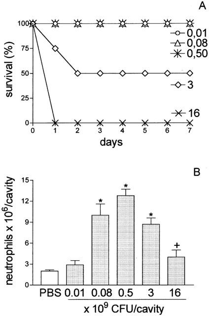
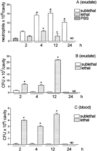
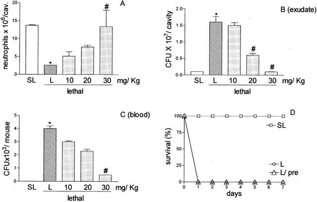
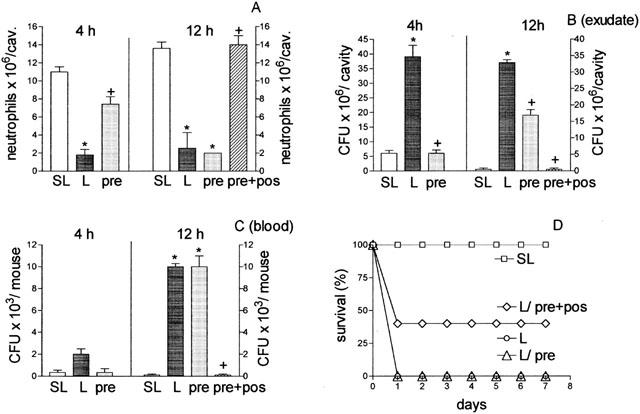
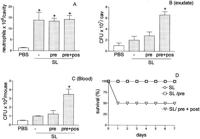
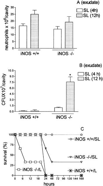
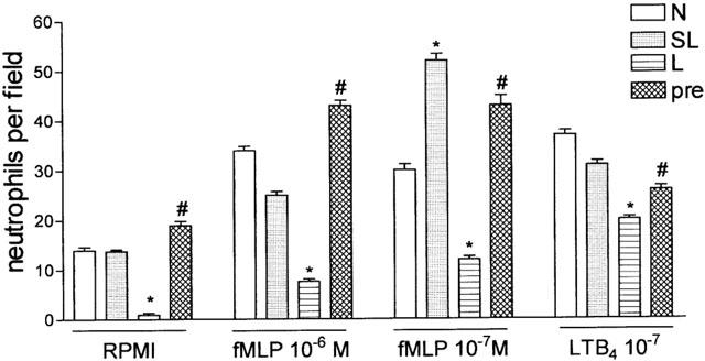
1. Sepsis induced by S. aureus was used to investigate whether neutrophil migration failure to infectious focus correlates with lethality in Gram-positive bacteria-induced sepsis in mice. 2. By contrast with the sub-lethal (SL-group), the lethal (L-group) intraperitoneal inoculum of S. aureus caused failure of neutrophil migration (92% reduction), high CFU in the exudate, bacteremia and impairment of in vitro neutrophil chemotactic activity. 3. Pre-treatments of L-group with adequate doses of aminoguanidine prevented the neutrophil migration failure and improved the survival of the animals (pre-treated: 43%; untreated: 0% survival). Thus, the impairment of neutrophil migration in the L-group appears to be mediated by nitric oxide (NO). 4. The injection of S. aureus SL-inoculum in iNOS deficient (-/-) or aminoguanidine-treated wild-type mice (pre- and post-treatment), which did not present neutrophil migration failure, paradoxically caused severe peritonitis and high mortality. This fact is explainable by the lack of NO dependent microbicidal activity in migrated neutrophils. 5. In conclusion, although the NO microbicidal mechanism is active in neutrophils, the failure of their migration to the infectious focus may be responsible for the severity and outcome of sepsis.


Figures

Figure 1
Figure 1
Survival curve and quantification of neutrophil migration into the peritoneal cavity after S. aureus inoculation. Groups of animals received different bacterial inoculum (0.01, 0.08, 0.5, 3, 16×109 CFU/cavity; 500 μl; i.p.). (A) Survival was determined daily until 7 days after bacterial inoculation. Results are expressed as per cent survival and are representative of three different experiments (P<0.001, Mantel-cox log rank test). (B) Neutrophil migration into the peritoneal cavity was quantified 4 h after bacterial inoculation or PBS injection. Results are expressed as mean±s.e. neutrophils per cavity and are representative of three independent experiments (n=4). *P<0.01 compared with control group (PBS). +P<0.001 compared with 0.5×10⁹ CFU/animal (analysis of variance, followed by Bonferroni's test).
Figure 2
Figure 2
Quantification of neutrophil migration into the peritoneal cavity (A), CFU in the exudate (B), and in the blood (C). Groups of animals received i.p. sublethal (0.5×10⁹ CFU in 500 μl cavity−1) or lethal (16×109 CFU in 500 μl cavity−1) bacterial inoculations. (A) Neutrophil migration into the peritoneal cavity was determined 2, 4, 12 and 24 h after bacterial inoculation. Results are expressed as mean±s.e. neutrophils per cavity. (B and C) The CFU was determined in the exudate and blood collected at the times indicated. The exudate and blood were diluted, spread-plated and incubated at 37°C, and the CFU were determined 18 h later. Results are expressed as CFU per cavity (exudate) or as CFU per ml of blood and are representative of three different experiments (n=3 – 4 per experiment). *P<0.01 compared with PBS group. +P<0.05 compared with sublethal group. (Analysis of variance, followed by Bonferroni's test).
Figure 3
Figure 3
Concentration of TNF-α (A and B), IL-1β (C and D), and IL-10 (E and F) in the peritoneal exudate and in sera of mice injected with PBS, sub-lethal or lethal inoculums of S. aureus pre-treated or not with aminoguanidine. The cytokine concentrations in peritoneal exudate (left panels) and in sera (right panels) were determined at 4 h after PBS injection or inoculation of sub-lethal (SL, 0.5×10⁹ CFU/animal), or lethal (L, 16×109 CFU/animal) inoculums of S. aureus pre-treated with PBS or with aminoguanidine (Ag, 30 mg Kg−1, 30 min before bacterial inoculation). Results are expressed as mean±s.e. nanograms of cytokine per millilitre of exudate or sera (n=4). *P<0.01 compared with PBS group. +P<0.05 compared with sublethal inoculum. #P<0.05 compared with lethal inoculum (analysis of variance, followed by Bonferroni's test).
Figure 4
Figure 4
Effect of aminoguanidine pre-treatment of mice inoculated with S. aureus on neutrophil migration into the peritoneal cavity, CFU in exudate and in the blood, and survival. Lethal (16×109 CFU/cavity) inoculated mice were pre-treated with PBS or with 10, 20 or 30 mg Kg−1 aminoguanidine, subcutaneously, 30 min before S. aureus inoculation. Animals inoculated with the sub-lethal dose (0.5×10⁹ CFU/cavity) (SL) were pre-treated with PBS. Neutrophil migration into the peritoneal cavity (A), CFU in exudate (B) and in the circulation (C) were determined 4 h after bacterial inoculation. Results are expressed as mean±s.e. of neutrophils per cavity, or of CFU per cavity (exudate) or CFU per ml of blood. Results are representative of three independent experiments (n=4). +P<0.01 compared with sublethal inoculation. #P<0.05 compared with lethal inoculation pre-treated with PBS (analysis of variance, followed by Bonferroni's test). In D survival is expressed as per cent survival and was determined daily for 7 days (P<0.001 Mantelcox log rang test).
Figure 5
Figure 5
Effect in mice injected with a lethal inoculum of S. aureus of different schemes of aminoguanidine treatment on neutrophil migration into the peritoneal cavity (A) CFU in exudate (B), and in the sera (C), and on the survival of the animals (D). Lethal (L, 16×109 CFU/cavity) inoculated mice were pre-treated with PBS or with 30 mg Kg−1 of aminoguanidine, subcutaneously, 30 min before bacterial inoculation (pre) or 30 mg Kg−1 aminoguanidine, subcutaneously, 30 min before bacterial inoculation and 5 mg Kg−1, 6 h after (pre+post). The mice inoculated with the sub-lethal dose (SL, 0.5×10⁹ CFU/cavity) were pre-treated with PBS. Neutrophil migration into the peritoneal cavity (A) CFU in exudate (B) and in the blood (C) were determined 4 and 12 h after bacterial inoculation. Results are expressed as mean±s.e.+P<0.001 compared with sublethal group. #P<0.05 compared with lethal group treated with PBS (analysis of variance, followed by Bonferroni's test). In D survival of the animals is expressed as per cent and was determined daily for 7 days for all experimental groups (P<0.001 Mantel-cox log rang test). Results are representative of two independent experiments (n=4).
Figure 6
Figure 6
Effect of aminoguanidine pre-treatment of mice injected with sub-lethal dose of S. aureus on neutrophil migration into the peritoneal cavity (A), CFU in the exudate (B) and in the blood (C), and on survival of the animals (D). Sub-lethal (0.5×10⁹ CFU cavity−1) (SL) inoculated mice were pre-treated with PBS (−) or 30 mg Kg−1 of aminoguanidine, subcutaneously, 30 min before bacterial inoculation (pre) or 30 mg Kg−1 aminoguanidine, subcutaneously, 30 min before and 2 h after bacterial inoculation (pre+post). Neutrophil migration into the peritoneal cavity (A), CFU in the exudate (B) and in blood (C) were determined 4 h after bacterial inoculation. Results are expressed as mean±s.e. and are representative of three independent experiments (n=4). *P<0.05 compared with animals pre-treated with PBS. +P<0.01 when compared with SL-group. #P<0.01 when compared with L-group after pre-treatment (pre) (B and C) (analysis of variance, followed by Bonferroni's test). In (D) survival is expressed as % and was determined daily for 7 days (P<0.001 Mantel-cox log rang test).
Figure 7
Figure 7
Quantification of neutrophil migration into the peritoneal cavity (A), CFU in the peritoneal exudate (B) and survival of iNOS Knockout mice (C) injected with sub-lethal or lethal inoculums of S. aureus. Groups of control animals (iNOS+/+) or iNOS−/− received a sub-lethal (SL, 0.5×10⁹ CFU/cavity) i.p. inoculation of S. aureus. In (A) neutrophil migration into the peritoneal cavity was quantified 4 (SL or L) and 12 h (SL 12 h) after bacterial inoculation. Results are expressed as mean±s.e. neutrophils per cavity. In (B) exudate was collected 4 or 12 h after bacterial injection and the CFU in the exudate were assessed by dilution and spread-plating followed by counting 18 h later. Results are expressed as mean±s.e. CFU per cavity (exudate). *P<0.01 compared with iNOS−/− SL-inoculated mice (analysis of variance, followed by Bonferroni's test). In (D) survival of the animals is expressed as per cent survival and was determined daily for 7 days.
Figure 8
Figure 8
In vitro migration of neutrophils obtained from normal, sub-lethal and lethal inoculated mice pre-treated with PBS or with aminoguanidine. Blood neutrophils were collected and purified from normal mice (N), sub-lethal (SL, 0.5×10⁹ CFU/animal), or lethal (L, 16×109 CFU/animal) inoculated mice pre-treated with PBS or with aminoguanidine (pre, 30 mg Kg−1, s.c., 30 min. before lethal inoculation). Neutrophils (5×10⁴/well) were assayed for fMLP- and LTB4-induced chemotaxis (see Methods). Results are expressed as mean±s.e. neutrophils per field and are representative of two independent experiments (n=4). *P<0.001 compared with sub-lethal inoculation. #P<0.001 compared with lethal inoculation pre-treated with PBS (analysis of variance, followed by Bonferroni's test).

References

    1. AJUEBOR M.N., VIRÀG I., FLOWER R.J., PERRETTI M., SZABO C. Role of inducible nitric oxide synthase in the regulation of neutrophil migration in zymosan-induced inflammation. Immunology. 1998;95:625–630. - PMC - PubMed
    1. BAKER C.C., CHAUDRY I.H., GAINES H.O., BAUE A.E. Evaluation of factors affecting mortality rate after sepsis in a murine caecal ligation and puncture model. Surgery. 1983;94:331–335. - PubMed
    1. BANNAN J., VISVANATHAN K., ZABRIESKIE J.B. Structure and function of streptococcal and staphylococcal superantigens in septic shock. Infect. Dis. Clin. North Am. 1999;13:387–396. - PubMed
    1. BANICK P.D., CHEN Q., XU Y.A., THOM S.R. Nitric oxide inhibits neutrophil β2 integrin function by inhibition of membrane-associated cyclic GMP synthesis. J. Cell. Physiol. 1997;172:12–24. - PubMed
    1. BENJAMIM C.F., FERREIRA S.H., CUNHA F.Q. Role of nitric oxide in the failure of neutrophil migration in sepsis. J. Infect. Dis. 2000;182:214–223. - PubMed

Publication types

MeSH terms