

Airway function, oedema, cell infiltration and nitric oxide generation in conscious ozone-exposed guinea-pigs: effects of dexamethasone and rolipram
- PMID: 12086983
- PMCID: PMC1573394
- DOI: 10.1038/sj.bjp.0704764
Airway function, oedema, cell infiltration and nitric oxide generation in conscious ozone-exposed guinea-pigs: effects of dexamethasone and rolipram
Abstract
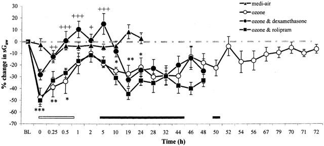
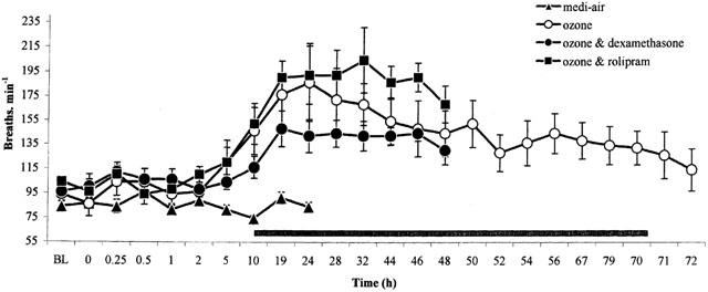
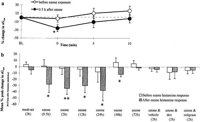
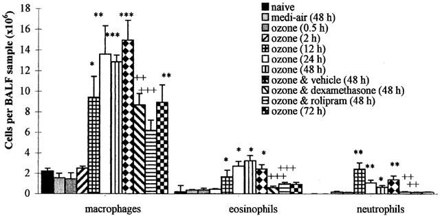
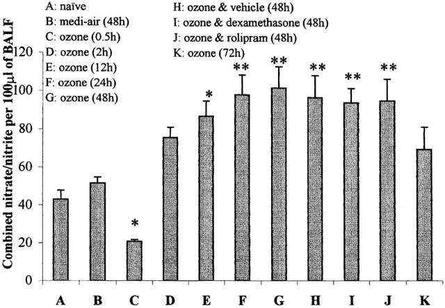
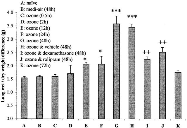
1. The effects of ozone inhalation (90 min, 2.15+/-0.05 p.p.m.) and their modification by dexamethasone (20 mg kg(-1)) or the phosphodiesterase-4 inhibitor, rolipram (1 mg kg(-1)), administered (i.p.) 24 and 0.5 h before and 24 h after ozone exposure were examined in conscious guinea-pigs. 2. Ozone caused an early-phase bronchoconstriction (EPB) as a fall in specific airways conductance (sG(aw)) measured by whole body plethysmography, followed at 5 h by a late-phase bronchoconstriction (LPB) and increased respiratory rate. Rolipram did not alter this profile but dexamethasone inhibited the EPB. 3. Airway hyperreactivity to inhaled histamine (1 mM, 20 s) occurred at 0.5, 2, 12, 24 and 48 h after ozone inhalation, the 2 h change being abolished by rolipram and dexamethasone. 4. Bronchoalveolar lavage fluid (BALF) macrophages, eosinophils and neutrophils were significantly (P<0.05) elevated at 12, 24 and 48 h after ozone exposure, the 48 h influx being significantly attenuated (P<0.05) by rolipram and dexamethasone. 5. BALF nitric oxide (NO) metabolites decreased 0.5 h after ozone exposure by 52%, recovered at 2 h and significantly increased at 12 (101%) and 24 h (127%). The elevated NO was unaffected by rolipram or dexamethasone. 6. Lung oedema, measured from wet/dry weight differences, was significant 12, 24 and 48 h after ozone exposure, the latter being significantly attenuated (P<0.05) by rolipram and dexamethasone. 7. Ozone exposure of guinea-pigs produced features common to COPD. Although rolipram and dexamethasone did not affect the airway function changes, they inhibited the inflammation, airway hyperreactivity and oedema.
Figures
Similar articles
-
Chronic lipopolysaccharide exposure on airway function, cell infiltration, and nitric oxide generation in conscious guinea pigs: effect of rolipram and dexamethasone.J Pharmacol Exp Ther. 2001 Jul;298(1):298-306. J Pharmacol Exp Ther. 2001. PMID: 11408555
-
Early and late bronchoconstrictions, airway hyper-reactivity, leucocyte influx and lung histamine and nitric oxide after inhaled antigen: effects of dexamethasone and rolipram.Clin Exp Allergy. 2004 Jan;34(1):91-102. doi: 10.1111/j.1365-2222.2004.01833.x. Clin Exp Allergy. 2004. PMID: 14720268
-
Airway function and reactivity, leukocyte influx and nitric oxide after inoculation with parainfluenza-3 virus: effects of dexamethasone or rolipram.Int Immunopharmacol. 2005 Apr;5(4):771-82. doi: 10.1016/j.intimp.2004.12.006. Int Immunopharmacol. 2005. PMID: 15710345
-
Elastolytic activity and alveolar epithelial type-1 cell damage after chronic LPS inhalation: effects of dexamethasone and rolipram.Toxicol Appl Pharmacol. 2005 Sep 15;207(3):257-65. doi: 10.1016/j.taap.2005.01.006. Toxicol Appl Pharmacol. 2005. PMID: 16129118
-
Goblet cell hyperplasia, airway function, and leukocyte infiltration after chronic lipopolysaccharide exposure in conscious Guinea pigs: effects of rolipram and dexamethasone.J Pharmacol Exp Ther. 2002 Aug;302(2):814-21. doi: 10.1124/jpet.102.033951. J Pharmacol Exp Ther. 2002. PMID: 12130748
Cited by
-
Role of mitogen-activated protein kinase phosphatase-1 in corticosteroid insensitivity of chronic oxidant lung injury.Eur J Pharmacol. 2014 Dec 5;744:108-14. doi: 10.1016/j.ejphar.2014.10.003. Epub 2014 Oct 12. Eur J Pharmacol. 2014. PMID: 25310910 Free PMC article.
-
Regulation of ozone-induced lung inflammation and injury by the β-galactoside-binding lectin galectin-3.Toxicol Appl Pharmacol. 2015 Apr 15;284(2):236-45. doi: 10.1016/j.taap.2015.02.002. Epub 2015 Feb 25. Toxicol Appl Pharmacol. 2015. PMID: 25724551 Free PMC article.
-
Inhibition of chlorine-induced pulmonary inflammation and edema by mometasone and budesonide.Toxicol Appl Pharmacol. 2013 Oct 15;272(2):408-13. doi: 10.1016/j.taap.2013.06.009. Epub 2013 Jun 22. Toxicol Appl Pharmacol. 2013. PMID: 23800689 Free PMC article.
-
Dexamethasone Inhibits Synergistic Induction of PDE4B Expression by Roflumilast and Bacterium NTHi.Int J Mol Sci. 2018 Nov 8;19(11):3511. doi: 10.3390/ijms19113511. Int J Mol Sci. 2018. PMID: 30413022 Free PMC article.
-
Lung macrophages: current understanding of their roles in Ozone-induced lung diseases.Crit Rev Toxicol. 2020 Apr;50(4):310-323. doi: 10.1080/10408444.2020.1762537. Epub 2020 May 27. Crit Rev Toxicol. 2020. PMID: 32458707 Free PMC article. Review.
References
-
- AGUSTI A.G.N., VILAVERDE J.M., TOGORES B., BOSCH M. Serial measurements of exhaled nitric oxide during exacerbations of chronic obstructive pulmonary disease. Eur. Respir. J. 1999;14:523–528. - PubMed
-
- ALVING K., WEITZBERG E., LUNDBERG J.M. Increased amount of nitric oxide in exhaled air of asthmatics. Eur. Respir. J. 1993;6:1368–1370. - PubMed
-
- ARAKIDA Y., OHGA K., SUWA K., OKADA Y., MORIO H., YOKOTA M., MIYATA K., YAMADA T., HONDA K. Effect of YM158, a dual lipid mediator antagonist, on immediate and late asthmatic responses, and on airway hyper-responsiveness in guinea pigs. Jpn. J. Pharmacol. 2000;82:287–294. - PubMed
-
- ARIS R.M., CHRISTIAN D., HEARNE P.Q., KERR K., FINKBEINER W.E., BALMES J.R. Ozone-induced airway inflammation in human subjects as determined by airway lavage and biopsy. Am. Rev. Respir. Dis. 1993;148:1363–1372. - PubMed
-
- BARNES P.J, , CHUNG K.F, PAGE C.P. Inflammatory mediators of asthma: an update. Pharmacol Rev. 1999;50:515–575. - PubMed
Publication types
MeSH terms
Substances
Grants and funding
LinkOut - more resources
Full Text Sources
Medical
