. 2002 Jul 9;99(14):9556-61.
doi: 10.1073/pnas.142298599. Epub 2002 Jun 26.

Role of the short isoform of myosin light chain kinase in the contraction of cultured smooth muscle cells as examined by its down-regulation

Affiliations

Role of the short isoform of myosin light chain kinase in the contraction of cultured smooth muscle cells as examined by its down-regulation

Jianjun Bao et al. Proc Natl Acad Sci U S A. .

Abstract

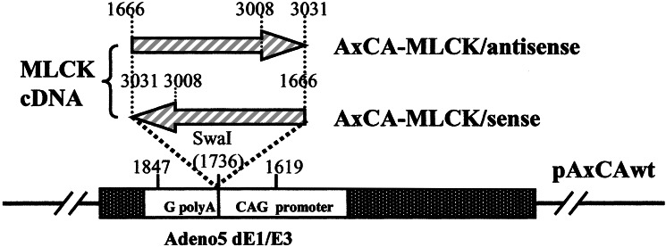
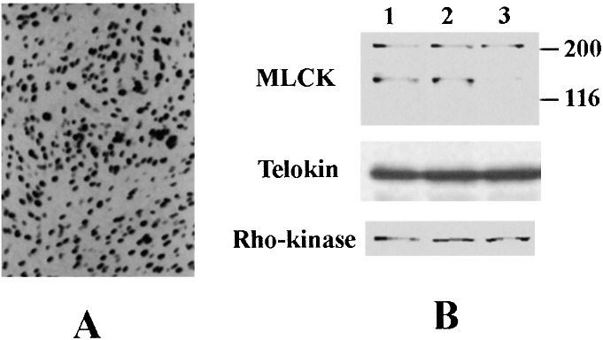
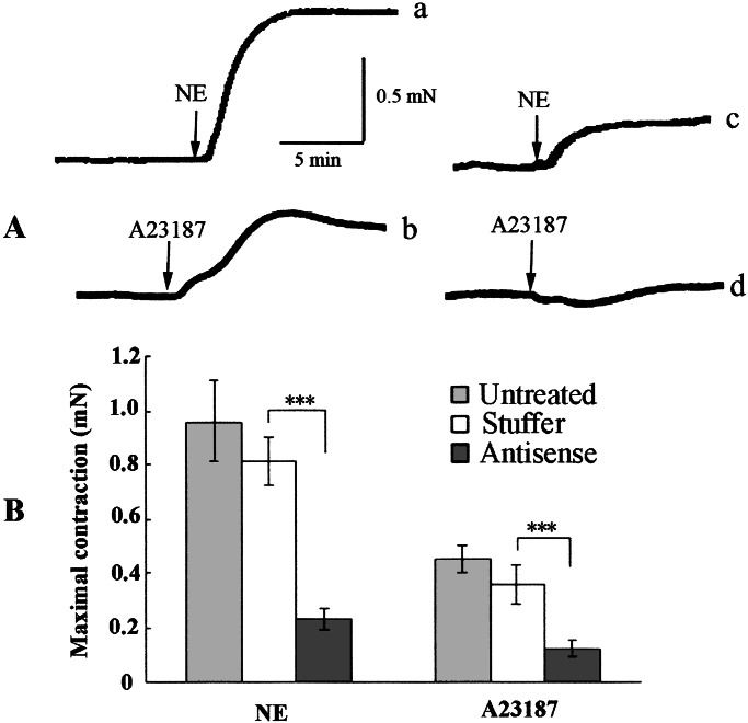
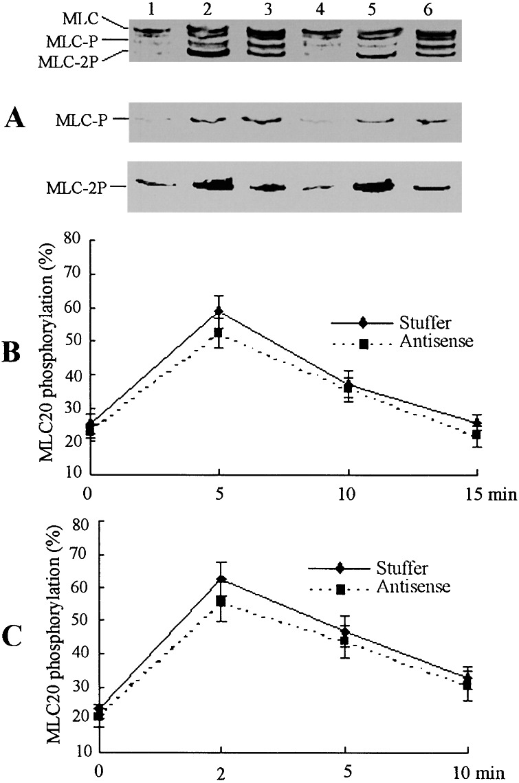
GbaSM-4 cells, smooth muscle cells derived from brain basilar artery, which express both 210-kDa long and 130-kDa short isoforms of myosin light chain kinase (MLCK), were infected with an adenovirus vector carrying a 1.4-kb catalytic portion of MLCK-cDNA in an antisense orientation. Western blot analysis showed that the expression of short MLCK was depressed without affecting long MLCK expression. The contraction of the down-regulated cells was measured by the cell-populated collagen-fiber method. The tension development after stimulation with norepinephrine or was depressed. The additional infection of the down-regulated cells with the adenovirus construct containing the same insert in a sense direction rescued not only the short MLCK expression but also contraction, confirming the physiological role of short MLCK in the contraction. To examine the role of long MLCK in the residual contraction persisting in the short MLCK-deficient cells, long MLCK was further down-regulated by increasing the multiplicity of infection of the antisense construct. The additional down-regulation of long MLCK expression, however, did not alter the residual contraction, ruling out the involvement of long MLCK in the contractile activity. Further, in the cells where short MLCK was down-regulated specifically, the extent of phosphorylation of 20-kDa myosin light chain (MLC20) after the agonist stimulation was not affected. This finding suggests that there are additional factors to MLC20 phosphorylation that contribute to regulate smooth muscle contraction.


Figures

Figure 1
Figure 1
Schematic representation of construction of the recombinant adenovirus vectors. We obtained adenoviral recombinants bearing a rabbit smooth muscle MLCK–cDNA fragment coding 1666–3031 bp in a sense or antisense orientation by COS/TPC method (18) and named then AxCA–MLCK/antisense and AxCA–MLCK/sense, respectively.
Figure 2
Figure 2
(A) Infection of GbaSM-4 with the adenovirus vectors. GbaSM-4 cells cultured in monolayers were infected with AxCA–NLacZ and incubated for 72 h. The transduction was verified by β-galactosidase staining. Original magnification, ×100. (B) Comparison of MLCK, telokin, and Rho-kinase expression in fibers reconstituted from GbaSM-4 cells by Western blot analyses. Each lane was loaded with 5 μg of protein and subjected to immunoblotting with monoclonal antibody against MLCK and polyclonal antibodies to Rho-kinase and telokin. Lane 1, Untreated GbaSM-4 cells; lane 2, GbaSM-4 cells infected with the control vector of AxCA-stuffer; lane 3, GbaSM-4 cells infected with the antisense vector of AxCA–MLCK/antisense; MLCK, MLCK expression; telokin, telokin expression; Rho-kinase, Rho-kinase expression.
Figure 3
Figure 3
(A) Typical record of contraction of cell-populated fibers. Fibers infected with AxCA-stuffer (traces a and c) or AxCA–MLCK/antisense (traces b and d) were contracted isometrically by stimulation with 10−7 M NE (traces a and b), or 5 × 10⁻⁶ M A23187 (traces c and d). (B) Means ± SE (n = 4) in mN of maximum tension are expressed by bars and error bars. ***, Significant at P < 0.001 level compared with AxCA-stuffer-transduced fibers.
Figure 4
Figure 4
Myosin light chain (MLC20) phosphorylation in reconstituted fibers. (A) AxCA-stuffer-transduced (lanes 1, 2, and 3) or AxCA–MLCK/antisense-transduced (lanes 4, 5, and 6) fibers were stimulated with NE 10−7 M (lanes 3 and 6) for 5 min or 5 × 10⁻⁶ M A23187 (lanes 2 and 4) for 2 min, then subjected to glycerol-PAGE and transferred to a nitrocellulose membrane. Unphosphorylated MLC20 (MLC), monophosphorylated MLC20 (MLC-P), and diphosphorylated MLC20 (MLC-2P) were simultaneously detected with monoclonal anti-MLC20 antiboby as shown in the Top. The blots were also reacted with anti-MLC-P antibody (Middle) and anti-MLC-2P antibody (Bottom) to detect Ser-19-monophosphorylated MLC20 and Thr-18/Ser-19-diphosphorylated MLC20 (15, 20). (B and C) Time course of MLC20 phosphorylation in fibers stimulated with 10−7 M NE (B) or 5 × 10⁻⁶ M A23187 (C). Values are means ± SE (n = 4).
Figure 5
Figure 5
Effects of ML-9 and Y-27632 on the contraction and on the MLC20 phosphorylation of smooth muscle fibers in which short MLCK was down-regulated. (A) AxCA–MLCK/antisense-transduced fibers were first contracted with 10−7 M NE (trace a). The fibers were washed for 60 min and incubated with 3 × 10⁻⁵ M ML-9 (trace b) or 1 × 10⁻⁶ M Y-27632 (trace c) for 20 min, then a second contraction was induced by the addition of 10−7 M NE. After another washing for 60 min, the third contraction was induced (trace d). (B) Means of the maximal contraction induced by 10−7 M NE (n = 4) was expressed by bars and error bars as means ± SE. ***, Significant at P < 0.01 level. (C) The extent of the MLC20 phosphorylation (%) was determined 5 min after the 10−7 M NE stimulation as described in the legend to Fig. 4. The bars and error bars express mean ± SE (n = 4). The double arrowheads and the arrowhead are the MLC20 phosphorylation before stimulation (see Fig. 4B) and after 10−7 M NE stimulation (see Fig. 4B), respectively. **, Significant at P < 0.01; ***, significant at P < 0.001.

Similar articles

Cited by

References

    1. Kamm K E, Stull J T. Annu Rev Pharmacol Toxicol. 1985;25:593–620. - PubMed
    1. Ye L-H, Hayakawa K, Kishi H, Imamura M, Nakamura A, Okagaki T, Takagi T, Iwata A, Tanaka T, Kohama K. J Biol Chem. 1997;272:32182–32189. - PubMed
    1. Smith L, Su X, Lin P-J, Zhi G, Stull J T. J Biol Chem. 1999;274:29433–29438. - PubMed
    1. Ito M, Dabrowska R, Guerriero V, Jr, Hartshorne D J. J Biol Chem. 1989;264:13971–13974. - PubMed
    1. Gallagher P J, Herring B P. J Biol Chem. 1991;266:23945–23952. - PMC - PubMed

Publication types

MeSH terms

LinkOut - more resources