. 2002 Jul 1;30(13):2817-24.
doi: 10.1093/nar/gkf392.

The mitochondrial DNA polymerase as a target of oxidative damage

Affiliations

The mitochondrial DNA polymerase as a target of oxidative damage

Maria A Graziewicz et al. Nucleic Acids Res. .

Abstract

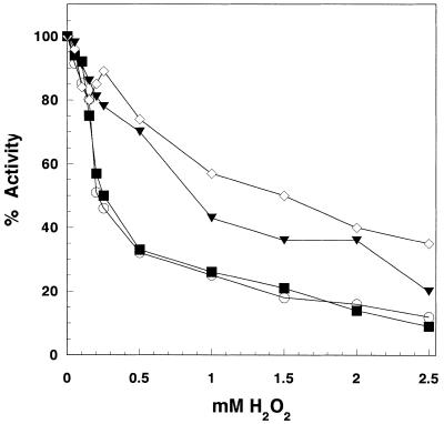
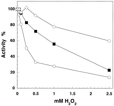
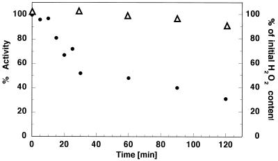
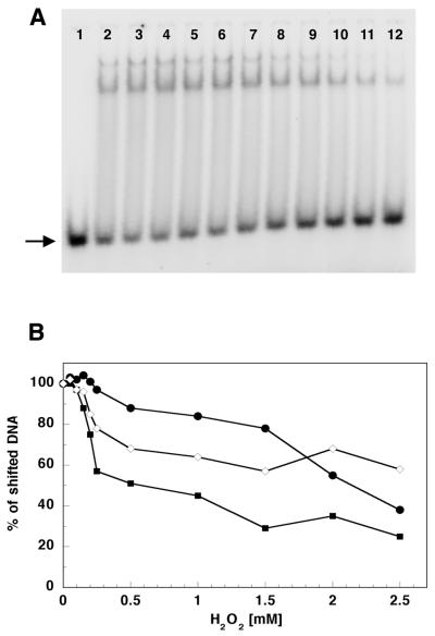
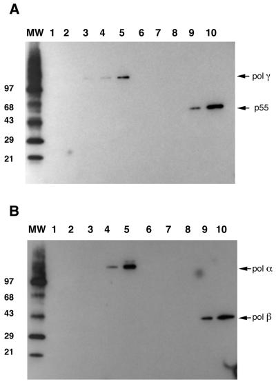
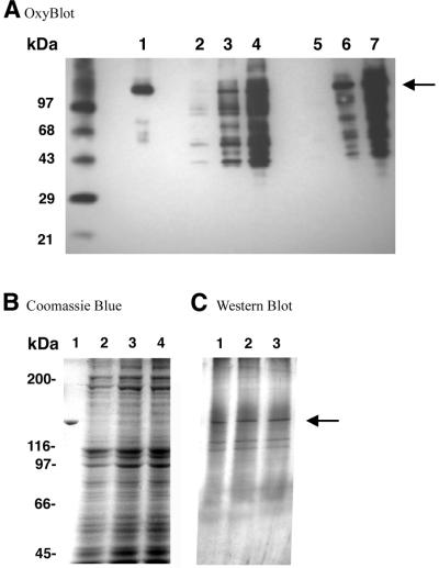
The mitochondrial respiratory chain is a source of reactive oxygen species (ROS) that are responsible for oxidative modification of biomolecules, including proteins. Due to its association with mitochondrial DNA, DNA polymerase gamma (pol gamma) is in an environment to be oxidized by hydrogen peroxide and hydroxyl radicals that may be generated in the presence of iron ions associated with DNA. We tested whether human pol gamma was a possible target of ROS with H2O2 and investigated the effect on the polymerase activities and DNA binding efficiency. A 1 h treatment with 250 microM H2O2 significantly inhibited DNA polymerase activity of the p140 subunit and lowered its DNA binding efficiency. Addition of p55 to the p140 catalytic subunit prior to H2O2 treatment offered protection from oxidative inactivation. Oxidatively modified amino acid residues in pol gamma resulting from H2O2 treatment were observed in vitro as well as in vivo, in SV40-transfected human fibroblasts. Pol gamma was detected as one of the major oxidized mitochondrial matrix proteins, with a detectable decline in polymerase activity. These results suggest pol gamma as a target of oxidative damage, which may result in a reduction in mitochondrial DNA replication and repair capacities.


Figures

Figure 1
Figure 1
Inhibition of pol γ DNA polymerase activity by hydrogen peroxide. Exop140 (0.2 pmol), either alone or with p55 (1:1 molar ratio), was preincubated for 60 min at 37°C with increasing concentrations of H2O2. Subsequently DNA polymerase activity on poly(dA)·oligo(dT) was measured as described in Materials and Methods. Open circles, Exop140; filled squares, Exop140 preincubated with H2O2 + p55; open diamonds, Exop140/p55 complex; filled triangles, Exop140 + p55 preincubated with H2O2 prior to adding to Exop140.
Figure 2
Figure 2
Effect of hydrogen peroxide on DNA polymerase activities of Exop140, pol β and pol α. All three enzymes were treated with H2O2 for 60 min and their DNA polymerase activities on poly(dA)·oligo(dT) were measured as described in Materials and Methods. Reaction mixtures contained ∼48 ng (0.4 pmol) Exop140 (open circles), 17 ng (0.4 pmol) pol β (open diamonds), ∼66 ng (0.4 pmol) pol α (filled squares), respectively.
Figure 3
Figure 3
Time course of pol γ catalytic subunit polymerase activity inhibition by 200 µM hydrogen peroxide. Exop140 was incubated either alone or with 200 µM H2O2 at 37°C. At the time intervals indicated samples were taken (0.2 pmol) and DNA polymerase activity (closed circles, left axis) on poly(dA)·oligo(dT) was measured as described in Materials and Methods. Percent activity at each time point was calculated relative to the control Exop140 samples collected at corresponding time points and used as the 100% reference. The rRight axis and open triangles depict the amount of H2O2 remaining after the incubation time relative to the zero time point.
Figure 4
Figure 4
Hydrogen peroxide affects the ability of p140, p55 and p140/p55 complex to bind to dsDNA. (A) An example of an autoradiogram of a gel mobility shift experiment showing p140 with no treatment (lane 2) or treatment with 0.05, 0.1, 0.15, 0.2, 0.25, 0.50, 1.0, 1.5, 2.0 and 2.5 mM H2O2 (lanes 3–12, respectively). Lane 1 contains the oligonucleotide substrate without addition of protein. Oligonucleotide substrate preparation and reaction conditions are described in Materials and Methods. Products were separated on a 15% polyacrylamide non-denaturating gel and quantitated on a Molecular Dynamics PhosphorImager as described in Materials and Methods. Two shifted products were observed corresponding to either one or two p140 molecules bound per oligonulceotide substrate. (B) DNA binding efficiency of p140 (filled squares), p55 (closed circles) and p140/p55 complex (open diamonds) after preincubation at increasing H2O2 concentrations. The arrow indicates the unshifted dsDNA substrate.
Figure 5
Figure 5
In vitro oxidation of Exop140, p55, pol β and pol α by hydrogen peroxide. Equal amounts of protein (6 µg) were incubated in the presence of increasing H2O2 concentrations as indicated. After 60 min incubation the excess H2O2 was removed by a 5 min incubation with catalase. Proteins were then separated by SDS–PAGE on 4–20% polyacrylamide gels, electrotransferred to an Imobilon P membrane (Millipore) and probed with anti-DNP antisera (Intergen) against derivatized carbonyl groups in oxidized proteins as described in Materials and Methods. (A) Oxidation of Expp140 and p55. Lanes 1–5 depict the oxidation of Exop140 with no treatment (lane 1) or treatment with 0.1, 0.5, 1 and 2.5 mM H2O2, respectively. Lanes 6–10 depict the oxidation of Hp55 with no treatment (lane 6) or treatment with 0.1, 0.5, 1 and 2.5 mM H2O2, respectively. (B) Oxidation of pol α and pol β. Lanes 1–5 depict the oxidation of pol α with no treatment (lane 1) or treatment with 0.1, 0.25, 0.5 and 1 mM H2O2, respectively. Lanes 6–10 depict the oxidation of pol β with no treatment (lane 6) or treatment with 0.1, 0.25, 0.5 and 1 mM H2O2, respectively. MW represents the oxidized molecular weight markers (Intergen) in kDa in both panels.
Figure 6
Figure 6
In vivo oxidation of pol γ catalytic subunit. (A) Crude mitochondrial lysates from SVNF cells treated with H2O2 were preincubated with DNP-hydrazine to derivatize carbonyl residues occurring in proteins as a result of their oxidation. After derivatization, polyclonal DPg antisera against the catalytic subunit of pol γ were used to immunoprecipitate p140. Both crude mitochondrial lysates and immunoprecipitates were resolved on 4–20% polyacrylamide gels and electrotransferred to an Imobilon P membrane. Derivatized moieties were then recognized by anti-DNP antibodies as described in Materials and Methods. MW, oxidized molecular weight markers; lane 1, 0.5 µg oxidized Exop140 as control; lanes 2–4, 150 µg protein from crude mitochondrial lysate from non-treated cells (lane 2) and from cells treated with 100 (lane 3) and 250 µM (lane 4) H2O2; lanes 5–7, immunoprecipitate from mitochondrial lysates of non-treated cells (lane 5) and from cells treated with 100 (lane 6) and 250 µM (lane 7) H2O2. (B) Coomassie Blue staining of the crude mitochondrial lysates represented in lanes 2–4 of (A). Lane 1, 0.5 µg Exop140 control; lanes 2–4, crude mitochondrial lysates (∼150 µg) from non-treated cells (lane 2) and from cells incubated with 100 (lane 3) and 250 µM (lane 4) H2O2. Protein molecular weight markers are indicated on the left. (C) Immunoblot utilizing polyclonal DPg antisera against the catalytic subunit of pol γ. Lanes 1–3, immunoprecipitates from mitochondrial lysates from non-treated cells (lane 1) and from cells treated with 100 (lane 2) and 250 µM (lane 3) H2O2. The arrow indicates the position of the pol γ catalytic subunit.

References

    1. Cortopassi G.A., Shibata,D., Soong,N.W. and Arnheim,N. (1992) A pattern of accumulation of a somatic deletion of mitochondrial DNA in aging human tissues. Proc. Natl Acad. Sci. USA, 89, 7370–7374. - PMC - PubMed
    1. Michikawa Y., Mazzucchelli,F., Bresolin,N., Scarlato,G. and Attardi,G. (1999) Aging-dependent large accumulation of point mutations in the human mtDNA control region for replication. Science, 286, 774–779. - PubMed
    1. Chance B., Sies,H. and Boveris,A. (1979) Hydroperoxide metabolism in mammalian organs. Physiol. Rev., 59, 527–605. - PubMed
    1. Ballinger S.W., Van Houten,B., Jin,G.F., Conklin,C.A. and Godley,B.F. (1999) Hydrogen peroxide causes significant mitochondrial DNA damage in human RPE cells. Exp. Eye Res., 68, 765–772. - PubMed
    1. Salazar J.J. and Van Houten,B. (1997) Preferential mitochondrial DNA injury caused by glucose oxidase as a steady generator of hydrogen peroxide in human fibroblasts. Mutat. Res., 385, 139–149. - PubMed

MeSH terms