

Case Reports
doi: 10.3348/kjr.2002.3.2.133.
Radiologic findings of multiple myeloma with gastric involvement: a case report
Affiliations
- PMID: 12087204
- PMCID: PMC2713837
- DOI: 10.3348/kjr.2002.3.2.133
Item in Clipboard
Case Reports
Radiologic findings of multiple myeloma with gastric involvement: a case report
Korean J Radiol.
2002 Apr-Jun.
Abstract
We report a case of multiple myeloma with gastric involvement occurring in a patient who underwent an upper gastrointestinal series (UGIS), CT and MRI. UGIS depicted a luminal protruding mass, while contrast-enhanced CT demonstrated marked thickening of the gastric wall, with subtle contrast enhancement. At T1- and T2-weighted MR imaging, the mass showed iso- and intermediate signal intensity, respectively. After the administration of contrast material, subtle homogeneous enhancement was apparent.
Figures
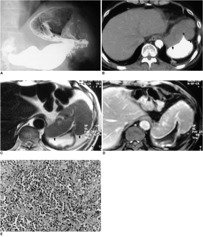
Multiple myeloma with gastric involvement in a 61-year-old man. A. Upper gastrointestinal series shows a large, protruding, luminal mass (arrows) with a well-defined margin in the anterior wall of the gastric cardia. B. Contrast-enhanced CT scan demonstrates marked gastric wall thickening with subtle contrast enhancement (arrows). C. Axial T2-weighted image shows homogeneous signal intensity slightly higher than that of the liver (arrows). D. Axial gadolinium-enhanced MR image obtained 3 minutes after the administration of contrast material depicts homogeneous enhancement, similar to that of hepatic parenchyma. E. Photograph of endoscopic biopsy specimen shows dense and monotonous infiltration by plasma cells (original magnification, ×200; hematoxylin-eosin staining). Note the presence of a monomorphic population of plasma cells with variable atypia.
Similar articles
-
Multiphasic CT and MRI appearances of extramedullary multiple myeloma involving the stomach, pancreas, and bladder.Clin Imaging. 2005 Jul-Aug;29(4):263-5. doi: 10.1016/j.clinimag.2004.11.002. Clin Imaging. 2005. PMID: 15967318
-
MR imaging of the gastrointestinal tract with i.v., gadolinium and diluted barium oral contrast media compared with unenhanced MR imaging and CT.AJR Am J Roentgenol. 1997 Oct;169(4):1051-9. doi: 10.2214/ajr.169.4.9308464. AJR Am J Roentgenol. 1997. PMID: 9308464
-
[A case of multiple myeloma presenting with a subcutaneous mass: significance of "dural tail sign" in the differential diagnosis of the meningeal tumors].No Shinkei Geka. 1999 Jan;27(1):67-71. No Shinkei Geka. 1999. PMID: 10024987 Japanese.
-
Role of MRI for the diagnosis and prognosis of multiple myeloma.Eur J Radiol. 2005 Jul;55(1):56-63. doi: 10.1016/j.ejrad.2005.01.017. Eur J Radiol. 2005. PMID: 15950101 Review.
-
Current role of CT in imaging of the stomach.Radiographics. 2003 Jan-Feb;23(1):75-87. doi: 10.1148/rg.231025071. Radiographics. 2003. PMID: 12533643 Review.
Cited by
-
Multiple Myeloma Presenting as Chronic Diarrhea.ACG Case Rep J. 2021 Nov 22;8(11):e00677. doi: 10.14309/crj.0000000000000677. eCollection 2021 Nov. ACG Case Rep J. 2021. PMID: 34820466 Free PMC article.
-
Multiple myeloma presenting as gastric polyposis.Dig Dis Sci. 2007 Dec;52(12):3340-2. doi: 10.1007/s10620-007-9775-7. Epub 2007 Apr 5. Dig Dis Sci. 2007. PMID: 17410432 No abstract available.
-
A Case of Concurrent Gastric and Pancreatic Plasmacytomas in a Patient With Multiple Myeloma: An Extremely Rare Entity.J Investig Med High Impact Case Rep. 2018 May 24;6:2324709618777003. doi: 10.1177/2324709618777003. eCollection 2018 Jan-Dec. J Investig Med High Impact Case Rep. 2018. PMID: 29854857 Free PMC article.
-
Extramedullary plasmacytoma mimicking colon carcinoma: an unusual presentation and review of the literature.BMJ Case Rep. 2015 Oct 23;2015:bcr2015210973. doi: 10.1136/bcr-2015-210973. BMJ Case Rep. 2015. PMID: 26498668 Free PMC article. Review.
-
A Durable Response of Primary Advanced Colonic Plasmacytoma Using a Combination of Surgical Resection and Adjuvant Bortezomib: A Case Report and Literature Review.Onco Targets Ther. 2022 Nov 7;15:1347-1354. doi: 10.2147/OTT.S372534. eCollection 2022. Onco Targets Ther. 2022. PMID: 36388154 Free PMC article.
References
-
- Kinoshita Y, Watanabe M, Takahashi H, et al. A case of gastric plasmacytoma: genetic analysis and immunofixation electrophoresis. Am J Gastroenterol. 1991;86:349–353. - PubMed
-
- Spagnoli I, Gattoni F, Mazzoni R, Uslenghi C. Primary gastrointestinal plasmacytoma: report of three cases. Diagn Imaging. 1983;52:23–27. - PubMed
-
- Yoon SE, Ha HK, Lee YS, et al. Upper gastrointestinal series and CT findings of primary gastric plasmacytoma: report of two cases. AJR. 1999;173:1266–1268. - PubMed
-
- Pimental RR, Van Stolk R. Gastric plasmacytoma: a rare cause of massive gastrointestinal bleeding. Am J Gastroenterol. 1993;88:1963–1964. - PubMed
-
- Remingio PA, Klaum A. Extramedullary plasmacytoma of the stomach. Cancer. 1971;27:562–568. - PubMed
Publication types
MeSH terms
Substances
LinkOut - more resources
Full Text Sources
Medical
