. 2002 Jun 5;86(11):1776-85.
doi: 10.1038/sj.bjc.6600329.

CD10 is a marker for cycling cells with propensity to apoptosis in childhood ALL

Affiliations
Free PMC article

CD10 is a marker for cycling cells with propensity to apoptosis in childhood ALL

G Cutrona et al. Br J Cancer. .
Free PMC article

Abstract

CD10 constitutes a favourable prognostic marker for childhood acute lymphoblastic leukaemia. Since correlations between CD10, cell cycle and apoptotic abilities were demonstrated in various cell types, we investigated whether differences existed in the cycling/apoptotic abilities of CD10-positive and CD10-negative B acute lymphoblastic leukaemia cells. Twenty-eight cases of childhood acute lymphoblastic leukaemia (mean age of 6.8 years) were subdivided into two groups according to high (17 cases, 93.2+/-4.5%, MRFI 211+/-82 CD10-positive cells) or low (11 cases, 11.5+/-6.2%, MRFI 10+/-7 CD10-negative cells) expression of CD10. CD10-positive acute lymphoblastic leukaemia cells were cycling cells with elevated c-myc levels and propensity to apoptosis, whereas CD10-negative acute lymphoblastic leukaemia cells had lower cycling capacities and c-myc levels, and were resistant to apoptosis in vitro. A close correlation between all these properties was demonstrated by the observations that the few CD10-positive cells found in the CD10-negative acute lymphoblastic leukaemia group displayed elevated c-myc and cycling capacities and were apoptosis prone. Moreover, exposure of CD10-positive acute lymphoblastic leukaemia B cells to a peptide nucleic acid anti-gene specific for the second exon of c-myc caused inhibition of c-myc expression and reduced cell cycling and apoptotic abilities as well as decreased CD10 expression.


Figures

Figure 1
Figure 1
Definition of CD10-positive and CD10-negative ALL. Bone marrow cell suspensions were stained for CD19 and CD10. CD19-positive cells were gated and the percentage of CD10-positive (top scale) cells or the MFRI for CD10 (bottom scale) were recorded. Two typical flow cytometry profiles for CD10 are also shown.
Figure 2
Figure 2
Apoptotic capacities of CD10-positive and CD10-negative ALL cells. Cells from the indicated cases were tested for apoptosis by PI staining either immediately after isolation or following 24 h in culture (left). Apoptotic capacities were confirmed by measuring DNA laddering (two typical experiments are shown on the right).
Figure 3
Figure 3
Cell cycle status of CD10-positive and CD10-negative ALL. (A and B) Cells from the indicated cases were stained for Ki67 (A) or BrdUrd (1 h pulse) (B) and analysed by flow cytometry. Typical profiles of CD10-positive or CD10-negative ALL cases are shown at the bottom of each figure. (C) Cells from a CD10-positive ALL were pulsed with BrdUrd for the indicated times, stained for PI and analysed by flow cytometry. Apoptotic (hypodiploid cells) are observed in the gates at the bottom of the figures.
Figure 4
Figure 4
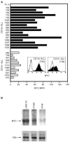
c-myc expression by ALL cells. (A) Cells from CD10-positive or CD10-negative ALL were stained for MYC protein following permeabilisation (see Materials and methods for details). The fluorescence detected by flow cytometry is expressed as MFRI. Two typical flow cytometry profiles are shown. (B) Western blot analysis of Myc expression by the cells from one CD10-positive (#1422) and one CD10-negative (#748) ALL case and by the BL cell line LAM C4. The Western blot was run with purified cell nuclei and probed with an anti Myc or an anti histone 2b antibody. This is a representative experiment of tests carried out on five CD10-positive and four CD10-negative ALL cases.
Figure 5
Figure 5
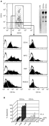
Features of CD10-positive cells isolated from CD10-negative ALL cases. (A) Cells from ALL case #571 were stained for CD10 and separated as indicated. Both fractions contained cells from the same clone as assessed by V(D)J length analysis (top right). Cells from the CD10-positive and CD10-negative cell fractions were stained for Myc and Ki67 or incubated with BrdUrd for 24 h before being stained with the appropriate reagents. The stained cells were analysed by flow cytometry. (B) The apoptotic capacities of CD10-positive and CD10-negative cell fractions purified from the indicated CD10-negative ALL cases were measured by PI staining before and after 24 h in culture.
Figure 6
Figure 6
Inhibition of c-myc expression by PNA-mycwt-NLS. (A) The indicated CD10-positive ALL were incubated with PNA-mycwt-NLS, PNA-mycmut-NLS or medium for 24 h and analysed for Myc expression by flow cytometry. (B and C) Cells from one representative CD10-positive ALL sample (#661) were cultured for 24 h and analysed for c-myc expression by RT–PCR (B) and Western blot (C). (C) Cells from one CD10-positive ALL case (#657) were treated with the indicated reagents for 24 h, pulsed with BrdUrd for 1 h and analysed by flow cytometry to determine the fraction of the cells in the S phase of the cell cycle.
Figure 7
Figure 7
Inhibition of apoptosis in CD10-positive ALL cells by PNA-mycwt-NLS. (A) Cells from the indicated cases were exposed to the indicated PNAs or to medium alone for 24 h in vitro. Apoptosis was then measured by PI or Annexin-V-FITC staining (not shown). (B) Apoptotis of cells from case #660 measured by PI or Annexin-V-staining. The cells were exposed to the indicated PNAs for 24 h in vitro prior to the test for apoptosis.
Figure 8
Figure 8
Inhibition of CD10 expression by PNA-mycmut-NLS. Cells from the indicated CD10-positive ALL cases were treated with PNAs or medium alone for 24 h in culture and subsequently stained for CD10. Fluorescence was measured by flow-cytometry and expressed as MFRI.

Similar articles

Cited by

References

    1. ArmitagePBerryG1987Statistical Methods in Medical Research,London: Blackwell Scientific Publications
    1. BahramFvon der LehrNCetinkayaCLarssonLG2000c-Myc hot spot mutations in lymphomas result in inefficient ubiquitination and decreased proteasome-mediated turnover Blood 1521042110 - PubMed
    1. BennettJCatovskyDDanielMTFlandrinGGaltonDAGralnickHRSultanC1981The morphological classification of acute lymphoblastic leukaemia: concordance among observers and clinical correlations Br J Haematol 47553561 - PubMed
    1. BissonnetteREcheverriFMahboubiAGreenDR1992Apoptotic cell death induced by c-myc is inhibited by bcl-2 Nature 359552554 - PubMed
    1. BorkhardtACazzanigaGViehmannSValsecchiMGLudwigWDBurciLMangioniSSchrappeMRiehmHLampertFBassoGMaseraGHarbottJBiondiA1997Incidence and clinical relevance of TEL/AML1 fusion genes in children with acute lymphoblastic leukemia enrolled in the German and Italian multicenter therapy trials. Associazione Italiana Ematologia Oncologia Pediatrica and the Berlin-Frankfurt-Munster Study Group Blood 90571577 - PubMed

Publication types

MeSH terms

LinkOut - more resources