. 2002 Jun 5;86(11):1803-12.
doi: 10.1038/sj.bjc.6600307.

Periodate-treated, non-anticoagulant heparin-carrying polystyrene (NAC-HCPS) affects angiogenesis and inhibits subcutaneous induced tumour growth and metastasis to the lung

Affiliations
Free PMC article

Periodate-treated, non-anticoagulant heparin-carrying polystyrene (NAC-HCPS) affects angiogenesis and inhibits subcutaneous induced tumour growth and metastasis to the lung

K Ono et al. Br J Cancer. .
Free PMC article

Abstract

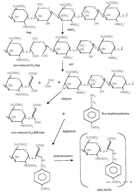
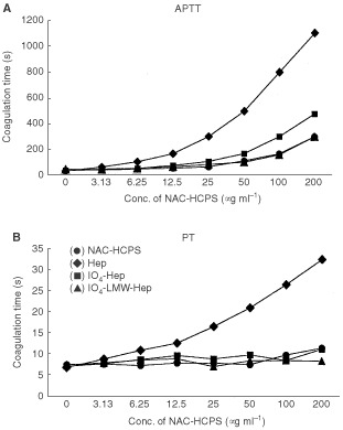
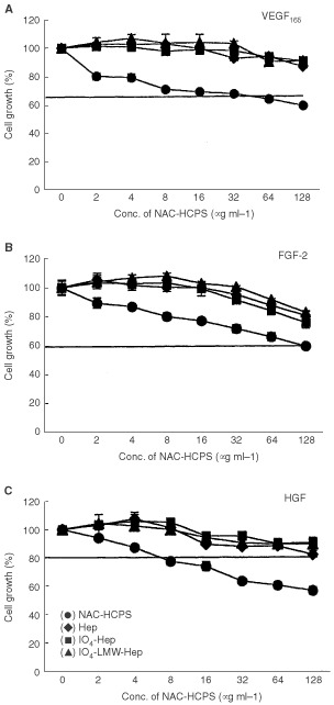
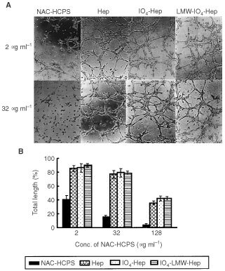
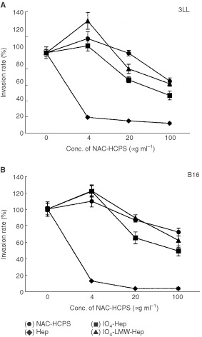
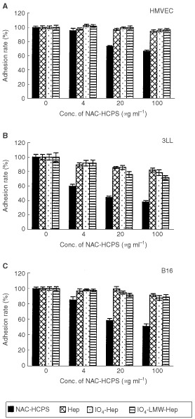
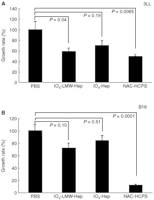
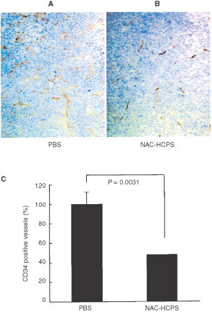
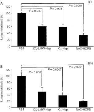
Periodate-treated, non-anticoagulant heparin-carrying polystyrene consists of about ten periodate-oxidized, alkaline-degraded low molecular weight-heparin chains linked to a polystyrene core and has a markedly lower anti-coagulant activity than heparin. In this study, we evaluated the effect of non-anticoagulant heparin-carrying polystyrene on tumour growth and metastasis. Non-anticoagulant heparin-carrying polystyrene has a higher activity to inhibit vascular endothelial growth factor-165-, fibroblast growth factor-2- or hepatocyte growth factor-induced human microvascular endothelial cell growth than heparin, ten periodate-oxidized-heparin and ten periodate-oxidized-low molecular weight-heparin, which is probably due to the heparin-clustering effect of non-anticoagulant heparin-carrying polystyrene. Non-anticoagulant heparin-carrying polystyrene inhibited human microvascular endothelial cell, B16 melanoma and Lewis lung cancer cell adhesion to Matrigel-coated plates. Non-anticoagulant heparin-carrying polystyrene also showed strong inhibitory activities in the tubular formation of endothelial cells on Matrigel and B16-melanoma and Lewis lung cancer cell invasion in a Matrigel-coated chamber assay. In vivo studies showed that growth of subcutaneous induced tumours and lung metastasis of B16-melanoma and Lewis lung cancer cells were more effectively inhibited by non-anticoagulant heparin-carrying polystyrene than ten periodate-oxidized-heparin and ten periodate-oxidized-low molecular weight-heparin. Furthermore, non-anticoagulant heparin-carrying polystyrene markedly reduced the number of CD34-positive vessels in subcutaneous Lewis lung cancer tumours, indicating a strong inhibition of angiogenesis. These results suggest that non-anticoagulant heparin-carrying polystyrene has an inhibitory activity on angiogenesis and tumour invasion and may be very useful in cancer therapy.


Figures

Figure 1
Figure 1
Reaction scheme for the preparation of IO4-Hep, IO4-LMW-Hep and NAC-HCPS.
Figure 2
Figure 2
Anticoagulant activity of NAC-HCPS. APTT (A) and PT (B) of rabbit plasma containing various concentrations of NAC-HCPS, Hep, IO4-Hep and IO4-LMW-Hep were measured, as has been described in Materials and Methods.
Figure 3
Figure 3
Effect of NAC-HCPS on growth factor-stimulated HMVEC growth. (A) VEGF165, (B) FGF-2, (C) HGF. Cell growth incubated with a growth factor in the absence of heparinoid was defined as 100% growth, and the data were calculated as a percentage. The horizontal line in each panel represents the level of cell growth obtained in the absence of both growth factor and heparinoid. The results represent the mean±s.e. in triplicate.
Figure 4
Figure 4
Effect of NAC-HCPS on fibroblast, 3LL and B16 cell growths. The cell growth in the absence of heparinoid was defined as 100% growth, and the data were calculated as a percentage. The results represent the mean±s.e. in triplicate.
Figure 5
Figure 5
Effect of NAC-HCPS on tubular formation of HMVEC. (A) Photomicrographs (original magnification: ×100) of the tubular formation of HMVEC cultured with 2 or 32 μg ml−1 of NAC-HCPS, Hep, IO4-Hep and IO4-LMW-Hep for 8 h on Matrigel-coated plates. Results are representatives of three independent experiments. (B) The quantitative evaluations of tubular formation of HMVEC cultured with various concentrations of NAC-HCPS, Hep, IO4-Hep and IO4-LMW-Hep.
Figure 6
Figure 6
Effect of NAC-HCPS on tumour cell invasion. Tumour cells (3LL (A) and B16 (B)) were seeded on a 8 μm pore size membrane coated with Matrigel. After 18 h incubation, invaded cells were stained and counted. The invasion rates were calculated as described in Materials and Methods. Results represent the mean±s.e. of four independent determinations.
Figure 7
Figure 7
Effect of NAC-HCPS on HMVEC, 3LL and B16 cell adhesions on Matrigel-coated plates. HMVEC, 3LL and B16 cells were plated on Matrigel-coated plates and incubated for 1 h. The bound cells were quantified as described in Materials and Methods. The results represent the mean±s.e. in triplicate.
Figure 8
Figure 8
Effect of NAC-HCPS on subcutaneous induced tumour growth of 3LL (A) and B16 (B) cells in mice. Tumour cells (1×10⁷) were implanted into the dorsal subcutis of mice. After tumours reached a measurable size (100∼200 mm3), 2 mg per 200 μl PBS of NAC-HCPS, IO4- Hep, IO4-LMW-Hep or PBS (200 μl) only was daily administered subcutaneously in the vicinity of the tumour for 7 days. Growth rates were calculated as described in Materials and Methods. Data were compared with the average tumour volume of the PBS treated group, defined as 100%.
Figure 9
Figure 9
Effect of NAC-HCPS on 3LL-tumour vascularisation. Vascularisation of the 3LL-tumour, evaluated immuno-histochemically with anti-murine CD34, markedly decreased in NAC-HCPS treated 3LL-tumours (B) when compared with PBS treated 3LL-tumours (A). The quantitative evaluation of the vascularisation (C) was carried out as described in Materials and Methods.
Figure 10
Figure 10
Effect of NAC-HCPS on lung colonisation of 3LL cells (A) and B16 cells (B) in mice. Both tumour cells (3×10⁵) were intraveneously injected through the lateral tail vein. From day 1 to day 7, either NAC-HCPS, IO4-Hep, IO4-LMW-Hep (1 mg per 100 μl of PBS) or 100 μl of PBS only was daily administered intraveneously through the lateral tail vein, and colony numbers on the lung surface in each mouse were counted on day 14.

References

    1. AlbiniAIwamotoYKleinmanHKMartinGRAarosonSAKozlowskiJMMcEwanRN1987A rapid in vitro assay for quantitating the invasion potential of tumour cells Cancer Res 4732393245 - PubMed
    1. BasbaumCBWerbZ1996Focalized proteolysis: Spatial and temporal regulation of extracellular matrix degradation at the cell surface Curr Opin Cell Biol 8731738 - PubMed
    1. BourinMCLindahlU1993Glycosaminoglycans and the regulation of blood coagulation Biochem J 289313330 - PMC - PubMed
    1. CollenASmorenburgSMPetersELupuFKoolwijkPVon NoordenCHinsberghVWM2000Unfractionated and low molecular weight heparin affect fibrin structure and angiogenesis in vitro Cancer Res 6061926200 - PubMed
    1. ConradHEGuoY1991Structural analysis of periodate-oxidized heparinInHeparin And Related Polysaccharides, Advances in Experimental Medicine and Biology 313Lane DA, Bjork I, Lindahl U (eds)pp3136New York: Plenum Publishing - PubMed

MeSH terms