. 2002 Jul;68(7):3597-605.
doi: 10.1128/AEM.68.7.3597-3605.2002.

Noninvasive quantitative measurement of bacterial growth in porous media under unsaturated-flow conditions

Affiliations

Noninvasive quantitative measurement of bacterial growth in porous media under unsaturated-flow conditions

R R Yarwood et al. Appl Environ Microbiol. 2002 Jul.

Abstract

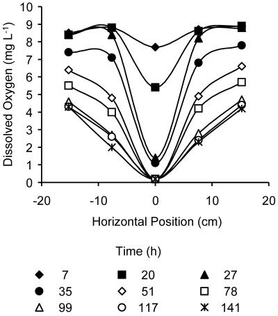
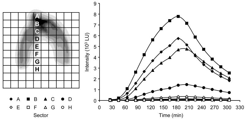
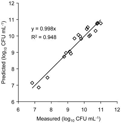
Glucose-dependent growth of the luxCDABE reporter bacterium Pseudomonas fluorescens HK44 was monitored noninvasively in quartz sand under unsaturated-flow conditions within a 45- by 56- by 1-cm two-dimensional light transmission chamber. The spatial and temporal development of growth were mapped daily over 7 days by quantifying salicylate-induced bioluminescence. A nonlinear model relating the rate of increase in light emission after salicylate exposure to microbial density successfully predicted growth over 4 orders of magnitude (r(2) = 0.95). Total model-predicted growth agreed with growth calculated from the mass balance of the system by using previously established growth parameters of HK44 (predicted, 1.2 x 10(12) cells; calculated, 1.7 x 10(12) cells). Colonization expanded in all directions from the inoculation region, including upward migration against the liquid flow. Both the daily rate of expansion of the colonized zone and the population density of the first day's growth in each newly colonized region remained relatively constant throughout the experiment. Nonetheless, substantial growth continued to occur on subsequent days in the older regions of the colonized zone. The proportion of daily potential growth that remained within the chamber declined progressively between days 2 and 7 (from 97 to 13%). A densely populated, anoxic region developed in the interior of the colonized zone even though the sand was unsaturated and fresh growth medium continued to flow through the colonized zone. These data illustrate the potential of a light transmission chamber, bioluminescent bacteria, and sensitive digital camera technology to noninvasively study real-time hydrology-microbiology interactions associated with unsaturated flow in porous media.


Figures

FIG. 1.
FIG. 1.
Temporal and spatial colonization of the light transmission chamber by P. fluorescens HK44 visualized by salicylate-inducible bioluminescence. The first panel shows a view of the chamber under room lighting. Drilled sampling port locations are marked by crosses. The superimposed rectangle defines the region of the chamber shown in each of the subsequent panels. The images have been artificially colorized to clearly show differences in emission intensity (arbitrary LU). The cross superimposed on each image indicates the location of the inoculation port, which is also shown as the uppermost port in the first panel. Digital image processing was carried out using Transform 3.4 (Fortner Software LLC), and processed images were prepared for publication using Photoshop 5.0 (Adobe Systems Inc., San Jose, Calif.).
FIG. 2.
FIG. 2.
Downstream dissolved oxygen dynamics in response to growth of P. fluorescens HK44 in the light transmission chamber. The horizontal axis indicates the position (distance to the left or right) of each sampling port with respect to the vertical center line of the chamber (defined as 0). The ports are marked as the horizontal row of crosses in the first panel in Fig. 1. The elapsed times after the start of the experiment are indicated.
FIG. 3.
FIG. 3.
Day 7 P. fluorescens HK44 bioluminescence response profiles. Light emission values represent the mean cumulative counts recorded for each 2.5- by 2.5-cm region (A to H) shown in the left panel. Time zero is the start of the salicylate pulse (0.1 g liter−1).
FIG. 4.
FIG. 4.
(A) Light emission profile of a horizontal transect through the P. fluorescens HK44-colonized zone at a depth of 15 cm (day 3). a, colonized region; b, light emission from the lantern effect. (B). Daily areal expansion of the colonized zone. Shaded regions (1 to 7) represent the newly colonized area on each day. The dashed line illustrates the apparent boundary of the colonized region on day 7 if uncorrected for the lantern effect.
FIG. 5.
FIG. 5.
Model-predicted and measured population densities of P. fluorescens HK44 associated with 2.5- by 2.5-cm sand samples (n = 19) recovered from bioluminescing regions of the colonized zone.

References

    1. Allison, D. G., B. Ruiz, C. SanJose, A. Jaspe, and P. Gilbert. 1998. Extracellular products as mediators of the formation and detachment of Pseudomonas fluorescens biofilms. FEMS Microbiol. Lett. 167:179-184. - PubMed
    1. Applegate, B. M., S. R. Kehrmeyer, and G. S. Sayler. 1998. A chromosomally based tod-luxCDABE whole-cell reporter for benzene, toluene, ethylbenzene, and xylene sensing. Appl. Environ. Microbiol. 64:2730-2735. - PMC - PubMed
    1. Auerbach, I. D., C. Sorensen, H. G. Hansma, and P. A. Holden. 2000. Physical morphology and surface properties of unsaturated Pseudomonas putida biofilms. J. Bacteriol. 182:3809-3815. - PMC - PubMed
    1. Blouin, K., S. G. Walker, J. Smit, and R. F. B. Turner. 1996. Characterization of in vivo reporter systems for gene expression and biosensor applications based on luxAB luciferase genes. Appl. Environ. Microbiol. 62:2013-2021. - PMC - PubMed
    1. Brink, R. H., P. Dubach, and D. L. Lynch. 1960. Measurement of carbohydrates in soil hydrolyzates with anthrone. Soil Sci. 89:157-166.

Publication types

LinkOut - more resources