Case Reports
. 2002 Jul;40(7):2370-80.
doi: 10.1128/JCM.40.7.2370-2380.2002.

Characterization of an unusual Mycobacterium: a possible missing link between Mycobacterium marinum and Mycobacterium ulcerans

Affiliations
Case Reports

Characterization of an unusual Mycobacterium: a possible missing link between Mycobacterium marinum and Mycobacterium ulcerans

K Chemlal et al. J Clin Microbiol. 2002 Jul.

Abstract

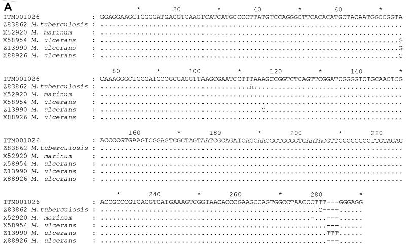
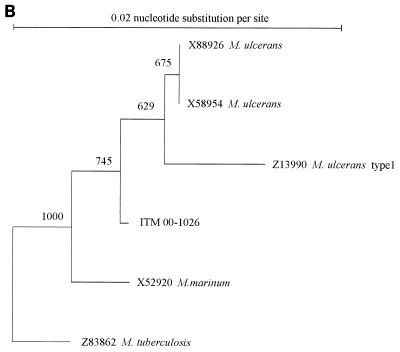
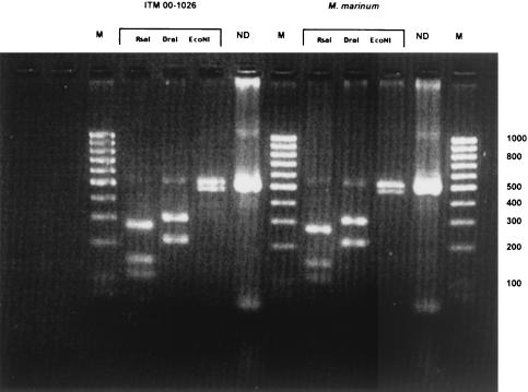
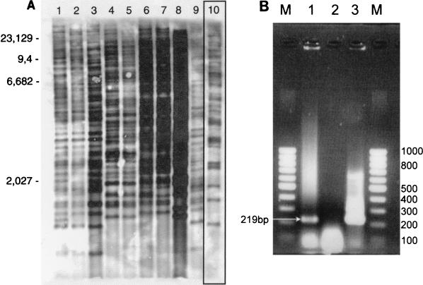
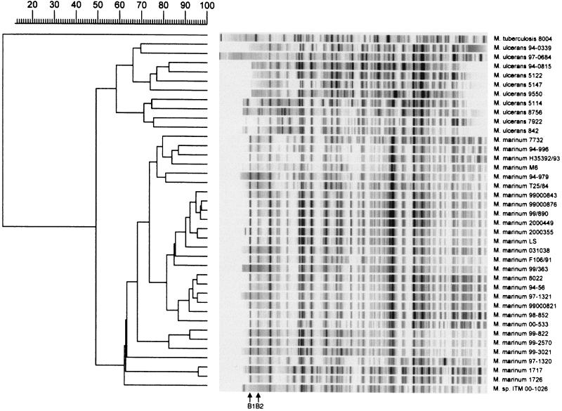
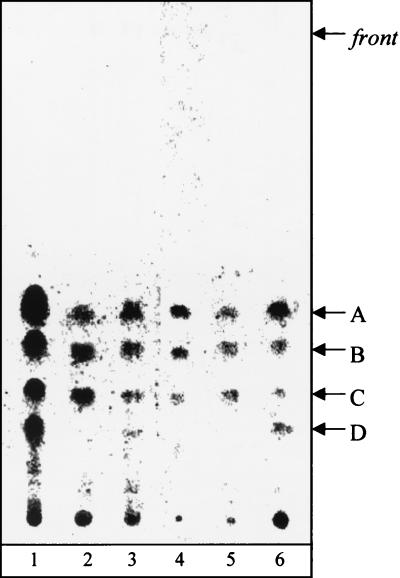
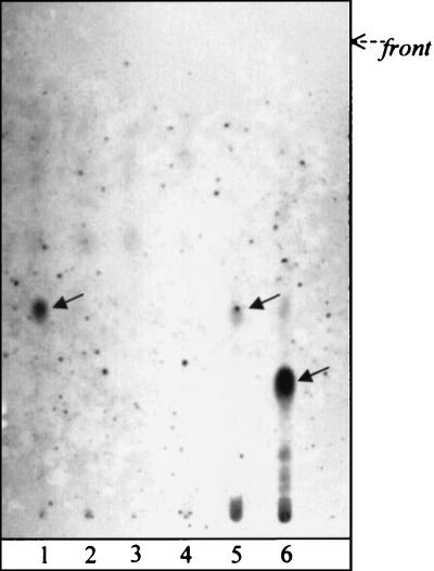
In an attempt to characterize an unusual mycobacterial isolate from a 44-year-old patient living in France, we applied phenotypic characterizations and various previously described molecular methods for the taxonomic classification of mycobacteria. The results of the investigations were compared to those obtained in a previous study with a set of temporally and geographically diverse Mycobacterium ulcerans (n = 29) and Mycobacterium marinum (n = 29) isolates (K. Chemlal, G. Huys, P.-A. Fonteyne, V. Vincent, A. G. Lopez, L. Rigouts, J. Swings, W. M. Meyers, and F. Portaels, J. Clin. Microbiol. 39:3272-3278, 2001). The isolate, designated ITM 00-1026 (IPP 2000-372), is closely related to M. marinum according to its phenotypic properties, lipid pattern, and partial 16S rRNA sequence. Moreover, fingerprinting by amplified fragment length polymorphism (AFLP) analysis unequivocally classified this strain as a member of the species M. marinum, although it lacked two species-specific AFLP marker bands. However, PCR and restriction fragment length polymorphism analysis based on M. ulcerans-specific insertion sequence IS2404 showed the presence of this element in a low copy number in isolate ITM 00-1026. In conclusion, the designation of this isolate as a transitional species further supports the recent claim by Stinear et al. (T. Stinear, G. Jenkin, P. D. Johnson, and J. K. Davies, J. Bacteriol. 182:6322-6330, 2000) that M. ulcerans represents a relatively recent phylogenetic derivative of M. marinum resulting from the systematic acquisition of foreign DNA fragments.


Figures

FIG. 1.
FIG. 1.
(A) Alignment of the 3′ end of the 16S rRNA sequence of ITM 00-1026 with selected closely related mycobacterial 16S rRNA. The numbering of the region in the 16S rRNA corresponds to that of the M. ulcerans sequence (base pairs 1110 to 1522) (GenBank reference sequence). Only nucleotides that differ from the reference sequence are shown. Dashes indicate deletions or absent nucleotides. (B) Phylogenetic tree based on the alignments of the partial 16S rRNA gene sequences illustrating the position of isolate ITM 00-1026 in relation to those of other closely related mycobacteria. The tree was constructed by using the neighbor-joining method. Bootstrapping (1,000 replicates) was used to assess support for particular nodes in the tree, and the values are shown above the nodes.
FIG. 1.
FIG. 1.
(A) Alignment of the 3′ end of the 16S rRNA sequence of ITM 00-1026 with selected closely related mycobacterial 16S rRNA. The numbering of the region in the 16S rRNA corresponds to that of the M. ulcerans sequence (base pairs 1110 to 1522) (GenBank reference sequence). Only nucleotides that differ from the reference sequence are shown. Dashes indicate deletions or absent nucleotides. (B) Phylogenetic tree based on the alignments of the partial 16S rRNA gene sequences illustrating the position of isolate ITM 00-1026 in relation to those of other closely related mycobacteria. The tree was constructed by using the neighbor-joining method. Bootstrapping (1,000 replicates) was used to assess support for particular nodes in the tree, and the values are shown above the nodes.
FIG. 2.
FIG. 2.
PCR restriction profiles obtained for ITM 00-1026 and M. marinum with three restriction enzymes (RsaI, DraI, and EcoNI). Lanes M, 100-bp DNA ladder; lanes ND, no digested PCR product. The numbers on the right of the gel are molecular sizes (in base pairs).
FIG. 3.
FIG. 3.
(A) Representative Southern blot obtained with nine M. ulcerans isolates (lanes 1 to 9, respectively) from different geographic areas and isolate ITM 00-1026. Lane 1, reference strain ATCC 19423; lane 2, an isolate from the Democratic Republic of Congo; lane 3, an isolate from Papua New Guinea; lane 4, an isolate from China; lane 5, an isolate from Japan; lane 6, an isolate from Suriname; lane 7, an isolate from French Guiana; lane 8, an isolate from Mexico; lane 9, an isolate from Benin; lane 10, isolate ITM 00-1026. Note the low copy number of IS 2404 bands for isolte ITM 00-1026 compared to the numbers for the M. ulcerans isolates. Molecular sizes (in kilobases) are shown on the left. (B) Image of the amplification product (219 bp) from the culture of ITM 00-1026 obtained by PCR with primers specific for IS2404. Lane 1, ITM 00-1026; lane 2, negative control; lane 3, positive control (M. ulcerans); lanes M, molecular size marker (100-bp DNA ladder).
FIG. 4.
FIG. 4.
Numerical analysis of normalized AFLP bands pattern generated from M. ulcerans (n = 10) and M. marinum (n = 27) strains and from isolate ITM 00-1026 with the A02-T02 primer pair. The dendrogram was constructed by the unweighted pair group method with arithmetic averages, with correlation levels expressed as percentages of the Pearson product-moment correlation coefficient. B1 and B2, two AFLP marker bands specific to all M. marinum strains evaluated.
FIG. 5.
FIG. 5.
Thin-layer chromatography profiles of mycolates. Fatty acid methyl esters were from M. ulcerans ITM 5142 (lane 1), isolate ITM 00-1026 (lane 2), M. ulcerans ATCC 19423T (lane 3), M. tuberculosis H37Ra (ATCC 25177) (lane 4), M. marinum ATCC 927T (lane 5), and M. ulcerans ITM 00-1441(lane 6). A, α-mycolates; B, methoxymycolates; C, ketomycolates; D, phenolphthiodiolone diphthioceranates. The developing solvent was petroleum ether-diethyl ether (9:1 [vol/vol]; five runs). Visualization was done by spraying with molybdophosphoric acid (10% [wt/vol]) in ethanol and charring.
FIG. 6.
FIG. 6.
Thin-layer chromatography profiles of glycolipids. Lane 1, isolate ITM 00-1026; lane 2, M. ulcerans ATCC 19423T; lane 3, M. marinum ATCC 927T; lane 4, M. ulcerans 00-1441; lane 5, M. marinum CIPT 14012 0006; lane 6, M. tuberculosis Canetti. The developing solvent was CHCl3-CH3OH (95:5 [vol/vol]). Visualization was done by spraying with anthrone (0.2% [wt/vol] in H2SO4) and heating. Arrows indicate anthrone-positive spots corresponding to the phenolic glycolipids of isolate ITM 00-1026, M. marinum (mycoside G), and M. tuberculosis Canetti (PGL-Tb 1) in lanes 1, 5, and 6, respectively.
FIG. 7.
FIG. 7.
Matrix-assisted laser desorption ionization-time of flight mass spectra of lipids obtained after saponification of whole mycobacterial cells. The series of peaks between m/z 1118 and 1290 correspond to α-, methoxy-, and ketomycolates (26). In the higher masses, peaks corresponding to alkali-stable lipids were detected (phenolphthiodiolone diphthioceranate, m/z 1340, 1368, and 1396; phenol glycolipid, m/z 1528, 1556, and 1584). Samples were dissolved in chloroform (final concentration, 1 mM) and were applied to the sample plate as 1-μl droplets. 2,5-Dihydroxybenzoic acid was used as the matrix. The accelerating voltage was 20 kV in the positive mode.

References

    1. Aronson, J. D. 1926. Spontaneous tuberculosis in salt water fish. Infect. Dis. 39:315-320.
    1. Bhatt, A., and T. Kieser. 1999. Transposition of IS117 of Streptomyces coelicolor A3(2) in Mycobacterium smegmatis. Microbiology 145:1201-1207. - PubMed
    1. Chemlal, K., K. De Ridder, P. A. Fonteyne. W. M. Meyers, J. Swings, and F. Portaels. 2001. The use of IS2404 restriction fragment length polymorphism suggests the diversity of Mycobacterium ulcerans from different geographical areas. Am. J. Trop. Med. Hyg. 64:270-273. - PubMed
    1. Chemlal, K., G. Huys, P.-A. Fonteyne, V. Vincent, A. G. Lopez, L. Rigouts, J. Swings, W. M. Meyers, and F. Portaels. 2001. Evaluation of PCR restriction profile analysis, IS2404 restriction fragment length polymorphism and amplified fragment length polymorphism fingerprinting for identification and typing of Mycobacterium ulcerans and M. marinum. J. Clin. Microbiol. 39:3272-3278. - PMC - PubMed
    1. Daffé, M., M.-A. Lanéelle, and C. Lacave. 1991. Structure and stereochemistry of mycolic acids of Mycobacterium marinum and Mycobacterium ulcerans. Res. Microbiol. 142:397-403. - PubMed

Publication types

MeSH terms

LinkOut - more resources