

Detection and genotyping of human group A rotaviruses by oligonucleotide microarray hybridization
- PMID: 12089254
- PMCID: PMC120567
- DOI: 10.1128/JCM.40.7.2398-2407.2002
Detection and genotyping of human group A rotaviruses by oligonucleotide microarray hybridization
Abstract
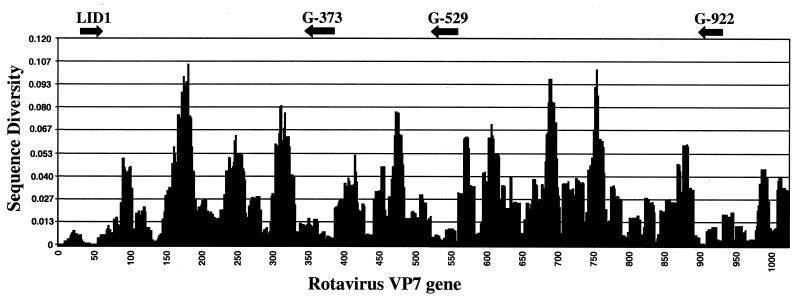
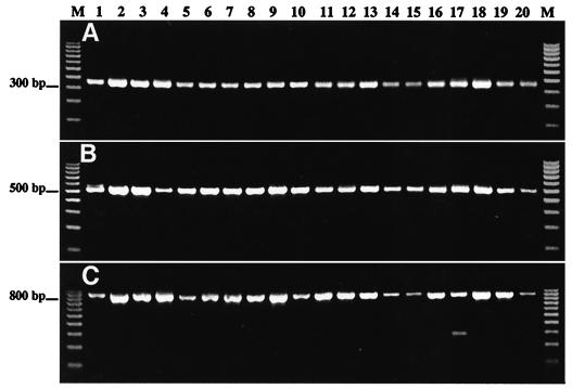
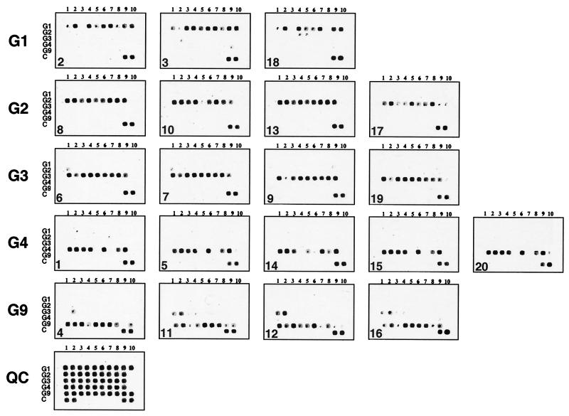
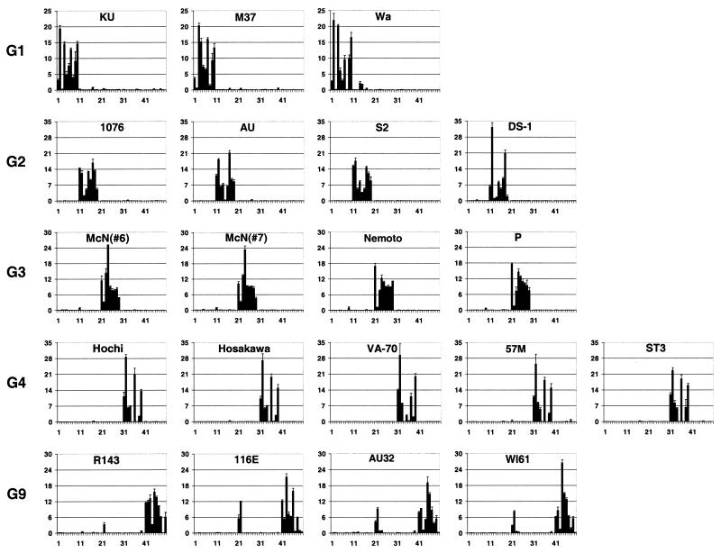
A rapid and reliable method for the identification of five clinically relevant G genotypes (G1 to G4 and G9) of human rotaviruses based on oligonucleotide microarray hybridization has been developed. The genotype-specific oligonucleotides immobilized on the surface of glass slides were selected to bind to the multiple target regions within the VP7 gene that are highly conserved among individual rotavirus genotypes. Rotavirus cDNA was amplified in a PCR with primers common to all group A rotaviruses. A second round of nested PCR amplification was performed in the presence of indodicarbocyanine-dCTP and another pair of degenerate primers also broadly specific for all genotypes. The use of one primer containing 5'-biotin allowed us to prepare fluorescently labeled single-stranded hybridization probe by binding of another strand to magnetic beads. The identification of rotavirus genotype was based on hybridization with several individual genotype-specific oligonucleotides. This approach combines the high sensitivity of PCR with the selectivity of DNA-DNA hybridization. The specificity of oligonucleotide microchip hybridization was evaluated by testing 20 coded rotavirus isolates from different geographic areas for which genotypes were previously determined by conventional methods. Analysis of the coded specimens showed that this microarray-based method is capable of unambiguous identification of all rotavirus strains. Because of the presence of random mutations, each individual virus isolate produced a unique hybridization pattern capable of distinguishing different isolates of the same genotype and, therefore, subgenotype differentiation. This strain information indicates one of several advantages that microarray technology has over conventional PCR techniques.
Figures
References
-
- Baldwin, D., V. Crane, and D. Rice. 1999. A comparison of gel-based, nylon filter and microarray techniques to detect differential RNA expression in plants. Curr. Opin. Plant Biol. 2:96-103. - PubMed
-
- Cheung, V. G., J. P. Gregg, K. J. Gogolin-Ewens, J. Bandong, C. A. Stanley, L. Baker, M. J. Higgins, N. J. Nowak, T. B. Shows, W. J. Ewens, S. F. Nelson, and R. S. Spielman. 1998. Linkage-disequilibrium mapping without genotyping. Nat. Genet. 18:225-230. - PubMed
Publication types
MeSH terms
Substances
LinkOut - more resources
Full Text Sources
Other Literature Sources
