. 2002 Jul;40(7):2545-54.
doi: 10.1128/JCM.40.7.2545-2554.2002.

Molecular peculiarities of the lytA gene isolated from clinical pneumococcal strains that are bile insoluble

Affiliations

Molecular peculiarities of the lytA gene isolated from clinical pneumococcal strains that are bile insoluble

Virginia Obregón et al. J Clin Microbiol. 2002 Jul.

Abstract

The autolytic LytA amidase from 12 bile (deoxycholate)-insoluble streptococcal isolates (formerly classified as atypical Streptococcus pneumoniae) showing different antibiotic resistance patterns was studied. These atypical strains, which autolyze at the end of the stationary phase of growth, contain highly divergent lytA alleles (pairwise evolutionary distances of about 20%) compared to the lytA alleles of typical pneumococci. The atypical LytA amidases exhibit a peculiar deletion of two amino acids responsible for cell wall anchoring in the carboxy-terminal domain and have a reduced specific activity. These enzymes were inhibited by 1% deoxycholate but were activated by 1% Triton X-100, a detergent that could be used as an alternative diagnostic test for this kind of strain. Preparation of functional chimeric enzymes, PCR mutagenesis, and gene replacements demonstrated that the characteristic bile insolubility of these atypical strains was due to their peculiar carboxy-terminal domain and that the 2-amino-acid deletion was responsible for the inhibitory effect of deoxycholate. However, the deletion alone did not affect the specific activity of LytA. A detailed characterization of the genes encoding the 16S rRNA and SodA together with multilocus sequence typing indicated that the strains studied here are not a single clone and, although they cannot be strictly classified as typical pneumococci, they represent a quite diverse pool of organisms closely related to S. pneumoniae. The clinical importance of these findings is underlined by the role of the lytA gene in shaping the course of pneumococcal diseases. This study can also contribute to solving diagnostic problems and to understanding the evolution and pathogenic potential of species of the Streptococcus mitis group.


Figures

FIG. 1.
FIG. 1.
Pairwise comparison of the nucleotide sequences of the lytA alleles from atypical isolates and from strain R6. Above the diagonal, a matrix of PEDs between aligned sequences is shown. Below the diagonal, the percent nucleotide identity is shown. Black, gray, and white boxes indicate identities lower than 85% (PEDs higher than 20%), between 90 and 97.5% (PEDs between 8 and 5%), and higher than 97.5% (PEDs lower than 2.5%), respectively.
FIG. 2.
FIG. 2.
Sequence variation in atypical lytA alleles. Each of the sites where the sequence of one or more of the lytA alleles differs from that of the wild type (lytAR6) is shown. Hyphens and colons represent nucleotides identical to those of the lytAR6 and lytA101 alleles, respectively. Sites where all of the sequences are identical are not shown. Sites 1, 2, and 3 indicate the first, second, and third nucleotide, respectively, in the codon. The numbering of the codons corresponds to that in a previous publication (16). Gray and black boxes indicate nucleotide changes causing conserved and nonconserved amino acid substitutions, respectively.
FIG. 3.
FIG. 3.
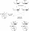
Schematic representation of the construction of chimeric genes between the lytA+ and lytA1338 alleles. The corresponding genes were PCR amplified with oligonucleotide primers LA5_Ext and LA3_Ext. The locations of the tandem promoters lppp-5 and lacpo in pINIII-A3 are shown. Abbreviations: bla, gene encoding β-lactamase; B, BamHI; H, HincII; X, XbaI. The elements of the figure are not drawn at the same scale.
FIG. 4.
FIG. 4.
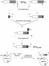
Construction of the expression vector pMVO1, encoding a 2-amino-acid deletion in the R6 amidase. The deleted motif is shown as a black box. The locations and directions of the oligonucleotide primers LA5_Ext (gray arrowhead), LA3_Ext (black arrowhead), MUTD1 (gray triangle), and MUTD2 (black triangle) are indicated. Abbreviations: B, BamHI; X, XbaI.
FIG. 5.
FIG. 5.
Multiple alignment of a partial nucleotide sequence of the lytA alleles from transformants MVO2, MVO4, and MVO5. The nucleotide sequences of the lytA alleles from strains 1338 (donor) and M41 (recipient) are also shown. Hyphens indicate nucleotides identical to those of the lytAR6 allele. Gray, stippled, and hatched bars represent the regions of lytAMVO2, lytAMVO4, and lytAMVO5, respectively, where recombination took place during transformation. The location of the HincII site used for constructing lytAN1338 and lytAC1338 (see Fig. 3) is also shown.
FIG. 6.
FIG. 6.
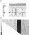
Sequence variation in sodA alleles. (A) Multiple alignment. Each of the sites where the sequence of one or more of the sodA alleles differs from that of the R6 strain is shown. Hyphens represent nucleotides identical to those of the sodAR6 allele. Sites where all of the sequences are identical are not shown. Sites 1, 2, and 3 indicate the first, second, and third nucleotide, respectively, in the codon. The numbering of the codons corresponds to that in a previous publication (53). Spn, Smi, and Sor indicate the sodA alleles from the type strains of S. pneumoniae, S. mitis, and S. oralis, respectively. (B) A matrix of PEDs between aligned sequences is shown. Numbers represent the estimated number of substitutions per 100 bases with no distance correction. Black boxes indicate comparisons with the sodA alleles from the type strains of S. pneumoniae (Spn), S. mitis (Smi), and S. oralis (Sor). The ranges of PEDs compared the atypical alleles with the sodA alleles included in the EMBL database (6 November 2001, last date accessed) and are shown in gray boxes. SPN, S. pneumoniae (26 entries); SMI, S. mitis (24 entries); SOR, S. oralis (40 entries).
FIG. 7.
FIG. 7.
Atypical LytA amidases contain a degenerate P6 motif. (A) Alignment of the C-terminal end of typical and atypical LytA amidases. The sequences of the bacterial LytA amidases were obtained from the EMBL database (10 December 2001, last date accessed). Eighteen typical and 19 atypical amidases, which include those reported here, were aligned by using pileup, and a consensus amino acid sequence of one enzyme of each class is shown. Colons indicate identical amino acid residues, and polymorphic residues are boxed. In the case of typical amidases, the consensus sequence contains amino acid residues present in at least 90% of the aligned enzymes, whereas a higher polymorphism was observed for atypical amidases. The corresponding accession numbers, positions, and changes in the atypical LytAs are: AJ252191, AJ252193, AJ419980, and AJ419981, 253G→R; AJ419974 and AJ419975, 253G→T; AJ252190, 253G→A; AJ419978 and AJ419982, 271L→P; S43511, 281N→H; AJ419977, 282A→V; AJ419978, 298T→P; S43511, AJ252191, AJ252192, AJ252193, AJ419977, and AJ419981, 301E→D; S43511, AJ252191, AJ252193, AJ419977, AJ419980, and AJ419981, 302K→R; S43511 and AJ419977, 306/307TV→SI; S43511 and AJ419977, 310E→D; S43511, 315V→M; AJ419976, 316K→I; and AJ419974, 316K→N. Choline-binding residues are indicated with a black triangle. The portions of the sequence that form the first and second strands of the hairpins are marked with an arrow. (B) Predicted three-dimensional folding of the P5 to P7 motifs of an typical LytA amidase lacking two amino acid residues (TG) at positions 290 to 291 (illustration 1) and of the corresponding consensus sequence from an atypical isolate (illustration 2). For simplicity, only the α-carbon chains are shown. The blue lines correspond to the modified amidases, whereas the folding of a typical LytA enzyme that has been experimentally determined (12) is drawn in yellow. Three different rotations of the models are shown.

Similar articles

Cited by

References

    1. Baba, T., and O. Schneewind. 1998. Targeting of muralytic enzymes to the cell division site of Gram-positive bacteria: repeat domains direct autolysin to the equatorial surface ring of Staphylococcus aureus. EMBO J. 17:4639-4646. - PMC - PubMed
    1. Denys, G. A., and R. B. Carey. 1992. Identification of Streptococcus pneumoniae with a DNA probe. J. Clin. Microbiol. 30:2725-2727. - PMC - PubMed
    1. Devereux, J., P. Haeberli, and O. Smithies. 1984. A comprehensive set of sequence analysis programs for the VAX. Nucleic Acids Res. 12:387-395. - PMC - PubMed
    1. Díaz, E., R. López, and J. L. García. 1990. Chimeric phage-bacterial enzymes: a clue to the modular evolution of genes. Proc. Natl. Acad. Sci. USA 87:8125-8129. - PMC - PubMed
    1. Díaz, E., R. López, and J. L. García. 1992. EJ-1, a temperate bacteriophage of Streptococcus pneumoniae with a Myoviridae morphotype. J. Bacteriol. 174:5516-5525. - PMC - PubMed

Publication types

MeSH terms

Associated data

LinkOut - more resources