. 2002 May;200(5):457-65.
doi: 10.1046/j.1469-7580.2002.00050.x.

The functional anatomy of the human anterior talofibular ligament in relation to ankle sprains

Affiliations

The functional anatomy of the human anterior talofibular ligament in relation to ankle sprains

T Kumai et al. J Anat. 2002 May.

Abstract

The anterior talofibular ligament is the most commonly injured ligament in the ankle. Despite considerable interest in the clinical outcome of treatment protocols, we do not know whether the distinctive pattern of localization of the injuries relates to regional differences in the structure and molecular composition of the ligament. To address this issue, ligaments were examined by histology and immunohistochemistry. Differences in the structure of its two attachments (i.e. entheses) were evaluated with quantitative, morphometric techniques, and regional differences in the distribution of collagens, glycosaminoglycans and proteoglycans were determined qualitatively by immunolabelling. Morphometric analyses showed that bone density was less at the fibular attachment, but that enthesis fibrocartilage was more prominent. Immunohistochemistry revealed the presence of a fibrocartilage (containing type II collagen and aggrecan) at the site where the ligament wraps around the lateral talar articular cartilage in a plantarflexed and inverted foot: the fibrocartilage is regarded as an adaptation to resisting compression. We propose that avulsion fractures are less common at the talar end of the ligament because (1) bone density is greater here than at the fibular enthesis, and (2) stress is dissipated away from the talar enthesis by the 'wrap-around' fibrocartilaginous character of the ligament near the talar articular facet.


Figures
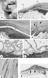
Fig. 1
Fig. 1
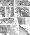
Basic structure of the ATFL. (a) A dissection of the ligament showing the region (arrow) where it wraps around the lateral articular facet of the talus (T). LM, lateral malleolus. (b) A low-power view of the talar end of the ATFL showing the presence of (b) A low-power view of the talar end of the ATFL showing the presence of a metachromatic, ‘sesamoid’ fibrocartilage (SF) in the region where the ligament contacts the articular cartilage (AC) on the talus. EF, enthesis fibrocartilage. Toluidine blue. Scale bar = 500 µm.(c) Enlargement of the region enclosed in the rectangle in b. Note the strong metachromasia of the sesamoid fibrocartilage (SF) and the presence of cartilage cell clusters (arrows) in both the ligament and the talar articular cartilage (AC). Toluidine blue. Scale bar = 100 µm. (d) The fibular enthesis of the ATFL showing the typical zones of uncalcified fibrocartilage (UF), calcified fibrocartilage (CF) and bone (B). Note that UF contains rows of fibrocartilage cells (arrows) and that it is separated from CF by a tidemark (TM). Toluidine blue. Scale bar = 100 µm. (e,f) Typical differences in bone density between the talar (e) and fibular (f) entheses of the ATFL – there is more bone at the talar end of the ligament. Both photographs are taken from the same ligament and at the same magnification. B, bone. M, marrow. Toluidine blue. Scale bar = 200 µm. (g) Rows of fibrocartilage cells (arrows) in the zone of uncalcified fibrocartilage at the fibular enthesis. Toluidine blue. Scale bar = 30 µm. (h) A marked decrease in the density of bony trabeculae that occurs at a point some distance beneath the fibular attachment of the ligament (large arrows). Note also the alignment of trabeculae along the long axis of the ligament (small arrows). Masson’s trichrome. Scale bar = 1 mm.
Fig. 2
Fig. 2
Histopathology and immunohistochemistry of the ATFL. (a) A small cyst (arrow) in the talar articular cartilage in the region where the latter articulates with the sesamoid fibrocartilage. Toluidine blue. Scale bar = 50 μm. (b) A small bony nodule (arrow) lying between two tidemarks (TM) within the zone of calcified fibrocartilage (CF) at the fibular enthesis. Toluidine blue. Scale bar = 100 µm. (c) A longitudinal fissure (F) within the zone of uncalcified fibrocartilage at the fibular enthesis associated with areas of intensely metachromatic ECM (arrows). Toluidine blue. Scale bar = 100 µm. (d) Degeneration of the opposing surfaces of the sesamoid fibrocartilage (SF) and talar articular cartilage (AC). Toluidine blue. Scale bar = 100 µm. Inset – cartilage cell clusters that were seen both in the talar articular cartilage and in the sesamoid fibrocartilage of the ATFL. Toluidine blue. Scale bar = 50 µm. (e) A bony spur (S) in the superficial part of the fibular enthesis of the ATFL. Scale bar = 200 µm. (f) An isolated bony nodule (N) that may represent an avulsion fragment near the fibular end of the ATFL. Note the presence of a depression of corresponding size on the lateral malleolus (asterisk), the secondary fibrocartilage (FC) that has developed on the bony nodule, and the presence of blood vessels in the intervening tissue (arrows). AC, articular cartilage. Scale bar = 2 mm. (g) Enlargement of the region where the secondary fibrocartilage (FC) on the bony nodule articulates with the hyaline articular cartilage on the fibula (AC). Masson’s trichrome. Scale bar = 100 µm. (h) Immunohistochemical labelling for type II collagen in the zones of calcified (CF) and uncalcified (UF) fibrocartilage at the fibular enthesis. B, bone. Scale bar = 100 µm. (i) Immunohistochemical labelling for aggrecan in the uncalcified fibrocartilage (UF) of the fibular enthesis. CF, calcified fibrocartilage; B, bone. Scale bar = 100 µm.(j) Immunohistochemical labelling for aggrecan in the sesamoid fibrocartilage near the talar end of the ligament. Note the greater intensity of labelling near the articular surface and the presence of rounded fibrocartilage cells (arrows). Scale bar = 50 µm.

References

    1. Attarian DE, McCrackin HJ, Devito DE, McElhsney JH, Garrett WE. Biomechanical charactersitics of human ankle ligaments. Foot Ankle. 1985;6:54–58. - PubMed
    1. Benjamin M, Evans EJ, Rao RD, Findlay JA, Pemberton DJ. Quantitative differences in the histology of the attachment zones of the meniscal horns in the knee joint of man. J. Anat. 1991;177:127–134. - PMC - PubMed
    1. Benjamin M, Newell RLM, Evans EJ, Ralphs JR, Pemberton DJ. The structure of the insertions of the tendons of biceps brachii, triceps and brachialis in elderly dissecting room cadavers. J. Anat. 1992;180:327–332. - PMC - PubMed
    1. Benjamin M, Qin S, Ralphs JR. Fibrocartilage associated with human tendons and their pulleys. J. Anat. 1995;187:625–633. - PMC - PubMed
    1. Benjamin M, Ralphs JR. Functional and development anatomy of tendon and ligaments. In: Gordon SL, Blair SJ, Fine LJ, editors. In Repetitive Motion Disorders of the Upper Extremity. Rosemont, Illinois: American Academy of Orthopaedic Surgeons; 1995. pp. 185–203.

MeSH terms