Review
. 2002 Apr;200(4):377-90.
doi: 10.1046/j.1469-7580.2002.00039.x.

Comparison of a new pmp22 transgenic mouse line with other mouse models and human patients with CMT1A

Affiliations
Review

Comparison of a new pmp22 transgenic mouse line with other mouse models and human patients with CMT1A

A M Robertson et al. J Anat. 2002 Apr.

Abstract

Charcot-Marie-Tooth disease type 1A is a dominantly inherited demyelinating disorder of the peripheral nervous system. It is most frequently caused by overexpression of peripheral myelin protein 22 (PMP22), but is also caused by point mutations in the PMP22 gene. We describe a new transgenic mouse model (My41) carrying the mouse, rather than the human, pmp22 gene. The My41 strain has a severe phenotype consisting of unstable gait and weakness of the hind limbs that becomes obvious during the first 3 weeks of life. My41 mice have a shortened life span and breed poorly. Pathologically, My41 mice have a demyelinating peripheral neuropathy in which 75% of axons do not have a measurable amount of myelin. We compare the peripheral nerve pathology seen in My41 mice, which carry the mouse pmp22 gene, with previously described transgenic mice over-expressing the human PMP22 protein and Trembler-J (TrJ) mice which have a P16L substitution. We also look at the differences between CMT1A duplication patients, patients with the P16L mutation and their appropriate mouse models.


Figures

Fig. 1
Fig. 1
Truncation of the 800-kb YAC MY451 to generate the 310-kb YAC MY451δ6 which was used for microinjection.
Fig. 2
Fig. 2
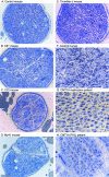
Histological sections. A,B,C,E: tibial nerves from 18-month-old mice. D: tibial nerve from 5-month-old mouse. F—H: sural nerves from patients.
Fig. 3
Fig. 3
Histograms of histological features in control, C61, C22, My41 and TrJ mice. Dysmyelinated fibres are given as the proportion of the total number of singly ensheathed fibres for six mice from each strain. Schwann cell nuclei are given as number counted per 100 axons from sciatic nerve for groups of six mice from each strain. g ratio is the ratio of axon diameter/fibre diameter for myelinated fibres from groups of six mice. Myelin periodicity (μm) was calculated for 20 fibres from each of six mice. Control, C61, C22 and TrJ mice (all 7–9 months), My41 mice (5 months old).
Fig. 4
Fig. 4
Fibre size distributions (μm) taken from the sciatic nerves of groups (n = 6) of Control, C61, C22 and TrJ mice (all 7–9 months old), and My41 mice (5 months old) and the sural nerve of representative control, CMT1A duplication and P16L patients. The percentage of myelinated or dysmyelinated fibres in each size range is indicated.
Fig. 5
Fig. 5
Unmyelinated fibre bundles. A: Remak fibres in a control mouse. B: extensive Schwann cell layers wrapping around Remak fibres in a C22 mouse. C: Schwann cell layers around Remak fibres in a TrJ mouse. D: layers of Schwann cell membrane seen around collagen pockets in a TrJ mouse.
Fig. 6
Fig. 6
Number of axons present in the mid sciatic nerves of 7–9-month-old animals (six of each strain), except My41 which were 5 months old (n = 6). Axons with a diameter greater than 1 μm were counted.
Fig. 7
Fig. 7
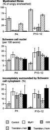
The development of myelination abnormalities in early postnatal mice. Graphs show the percentage of myelinated fibres, Schwann cell nuclei per 100 axons and the percentage of axons incompletely surrounded by Schwann cell cytoplasm. In each case, groups of seven mice (four in the case of My41) were counted. Animals were analysed at postnatal days 4 (P4) and postnatal days 10–12 (P10—12) as indicated.
Fig. 8
Fig. 8
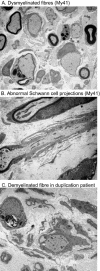
Schwann cell interactions. A: dysmyelinated axons incompletely surrounded by Schwann cell cytoplasm from a My41 mouse. B: abnormal Schwann cell cytoplasmic projections seen in this case in an My41 mouse. C: a demyelinated fibre from the sural nerve of a CMT1A duplication patient.
Fig. 10
Fig. 10
Schwann cell association with axons in humans and mice with the P16L mutation. Number of Schwann cell nuclei per 100 axons in three patients and six TrJ mice.

Similar articles

Cited by

References

    1. Ayers MM, Anderson RM. Onion bulb neuropathy in the Trembler mouse: a model of hypertrohic interstitial neuropathy (Dejerine-Sottas) in man. Acta. Neuropathol. 1973;25:54–70. - PubMed
    1. Baichwal RR, DeVries GH. A mitogen for Schwann cells derived from myelin basic protein. Biochem. Biophys. Res. Comm. 1989;164:883–888. - PubMed
    1. Berciano J, Garcia A, Calleja J, Combarros O. Clinico-electrophysiological correlation of extensor digitorum brevis muscle atrophy in children with Charcot-Marie-Tooth disease 1A duplication. Neuromusc. Disord. 2000;10:419–424. - PubMed
    1. Carenini S, Maurer M, Werner A, Blazyca H, Toyka KV, Schmid CD, et al. The role of macrophages in demyelinating peripheral nervous system of mice heterozygously deficient in pO. J. Cell. Biol. 2001;152:301–308. - PMC - PubMed
    1. Fullerton PM, Gilliatt RW. Pressure neuropathy in the hind foot of the guinea pig. J. Neurol. Neurosurg. Psychiatry. 1967;30:18–25. - PMC - PubMed

LinkOut - more resources