Review
. 2002 Apr;200(4):415-30.
doi: 10.1046/j.1469-7580.2002.00037.x.

Axons and glial interfaces: ultrastructural studies

Affiliations
Review

Axons and glial interfaces: ultrastructural studies

John Fraher. J Anat. 2002 Apr.

Abstract

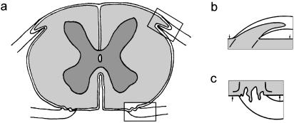
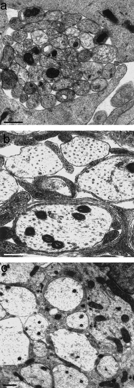
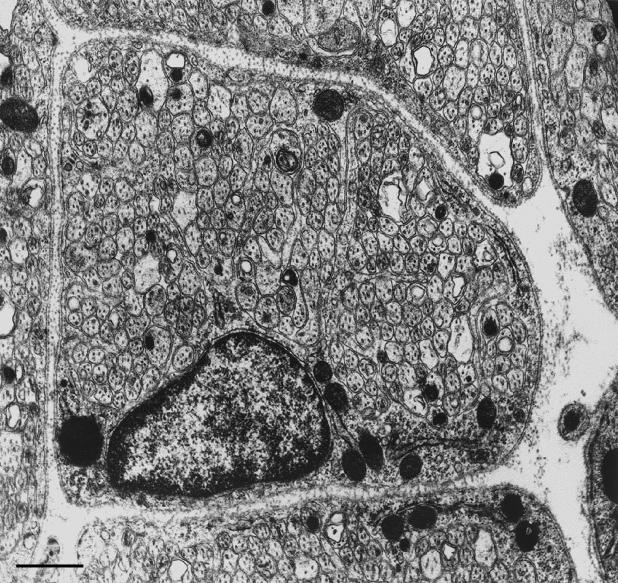
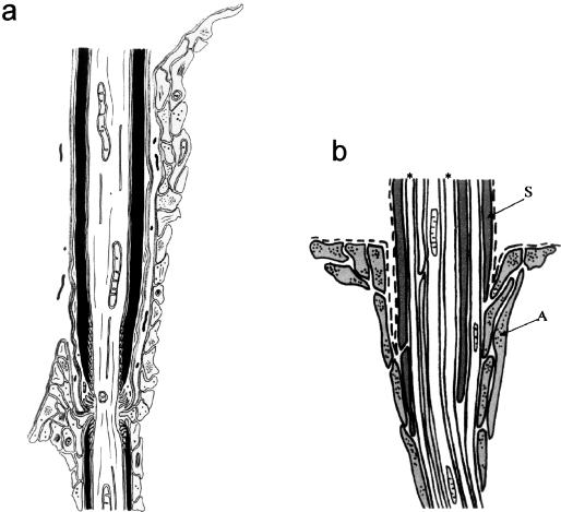
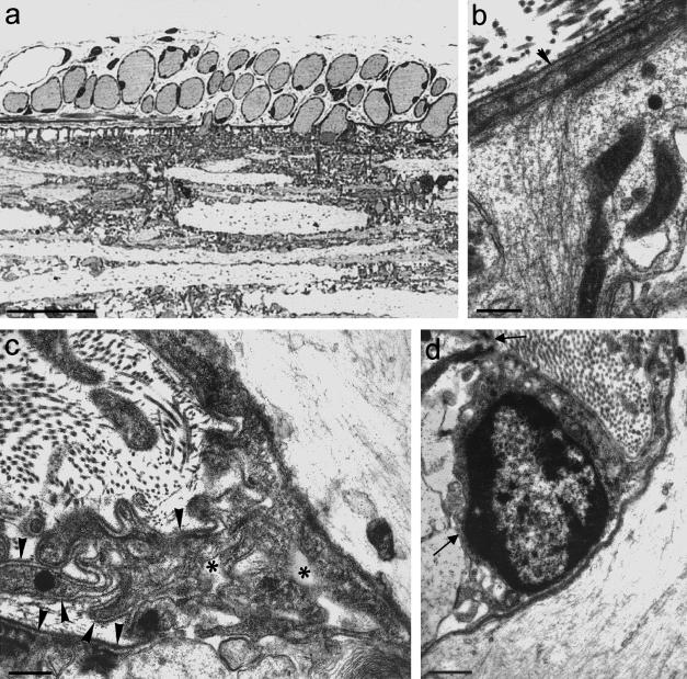
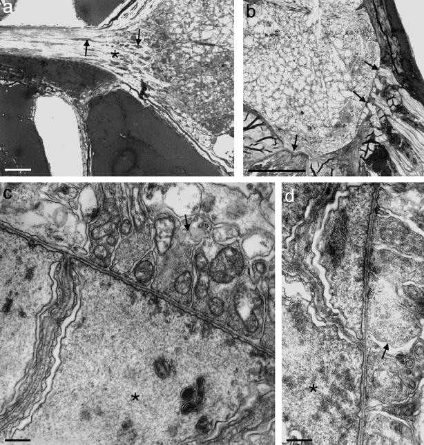
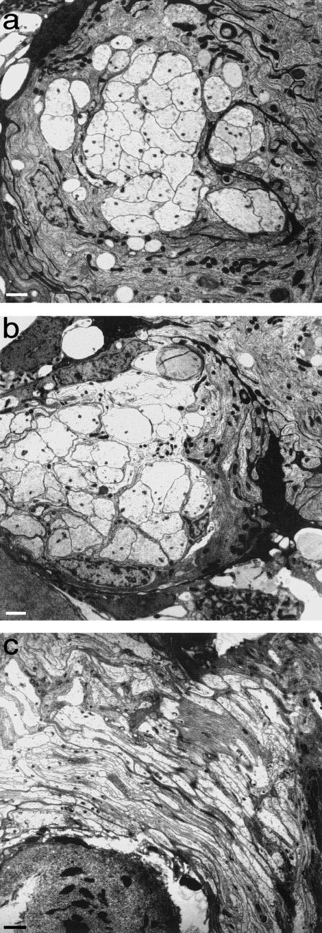
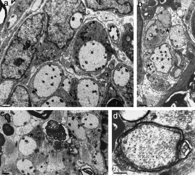
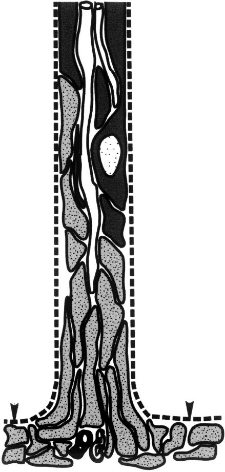
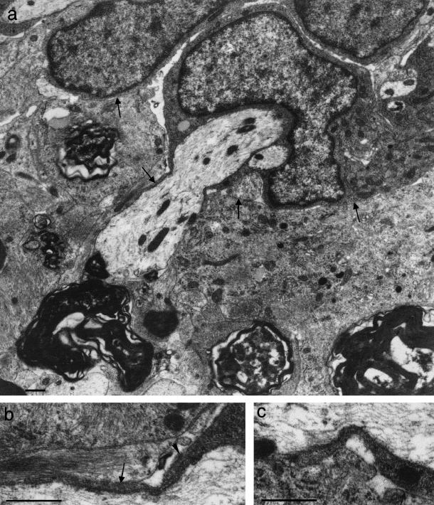
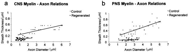
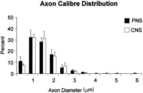
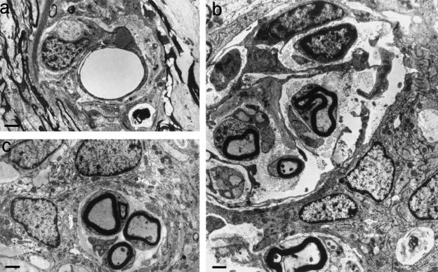
At most vertebrate nerve transitional zones (TZs) there is a glial barrier which is pierced by axons passing between the CNS and PNS. Myelinated axons traverse this in individual tunnels. The same is true of larger non-myelinated axons. This holds widely among the vertebrates, for example, the large motor axons of the sea-lamprey Petromyzon (which also possess TZ specializations not found in mammals). Smaller non-myelinated axons traverse the TZ glial tunnels as fascicles and so the barriers are correspondingly less comprehensive for them. Accordingly, in nerves composed of non-myelinated axons, such as the vomeronasal or the olfactory, a TZ barrier stretching across the nerve is effectivelyabsent. The chordateAmphioxus differsfrom the vertebrates in lacking a TZ barrier throughout. Invertebrates also lack glial barriers at the TZs between ganglia and interconnecting nerve trunks. The glial barrier at the dorsal spinal root TZ (DRTZ) has considerable value for analysing protocols aimed at achieving CNS regeneration, because it provides a useful model of the gliotic reaction at sites of CNS injury. Also, it is especially amenable to morphometric analysis, and so enables objective quantification of different protocols. Being adjacent to the subarachnoid space, it is accessible for experimental intervention. The DRTZ was used to investigate the value of neurotrophin 3 (NT3) in promoting axon regeneration across the TZ barrier and into the CNS following dorsal root crush. It promoted extensive regeneration and vigorous non-myelinated axonal ensheathment. On average, around 40% of regenerating axons grew across the interface, compared with virtually none in its absence. These may have traversed the interface through loci occupied by axons prior to degeneration. Many regenerating axons became myelinated, both centrally and peripherally.


Figures

Fig. 1
Fig. 1
Diagrams with enlargements of the areas outlined, showing (a) transversely sectioned spinal cord, (b) dorsal root attachment to the spinal cord, in which the rootlet contains a central tissue projection, (c) ventral root attachment to the spinal cord. Both show interdigitation of CNS and PNS tissues along the length of rootlet termed the transitional zone (TZ). Pale: PNS tissue; intermediate: CNS white matter; dark: CNS grey matter; Spinal cord surface: arrows.
Fig. 2
Fig. 2
At the developing TZ, presumptive large axons are (a) at first naked and emerge between loosely packed processes of the primitive glia limitans (above and below). (b,c) They become progressively segregated from one another by astrocytic processes which grow in from the surrounding cord surface (from Fraher, 1997). Scale bars: a: 1.0 μm; b,c: 0.5 μm.
Fig. 3
Fig. 3
Transversely sectioned vomeronasal nerve fascicle, showing it to be composed of large numbers of closely packed small axons, enveloped by a composite sleeve of ensheathing cells which segregate the axon bundle only to a limited extent. Scale bar: 0.5 μm.
Fig. 4
Fig. 4
Diagram showing the relationships between Schwann cells, astrocytes and axons at the TZ (a) in relation to a myelinated axon at the mature TZ and (b) in relation to a bundle of non-myelinated axons passing through the CNS–PNS interface (see text). (a) Astrocyte processes grey. (b) S: Schwann cell processes; A: astrocyte processes; dashed lines: basal lamina; asterisks: axons.
Fig. 5
Fig. 5
(a) Light micrograph of a longitudinal section of a lamprey spinal cord, showing transversely sectioned axons adjacent to, and traversing, the cord surface. Outside the cord each is surrounded by a thin, dark rim of Schwann cell cytoplasm. Scale bar: 50 μm. (b) Electron micrograph showing a dense layer of filaments in a surface process of the glia limitans (above), which extend deeply into its radially orientated extension. (Arrowhead: basal lamina) (from Fraher & Cheong, 1995). Scale bar: 0.25 μm. (c) Electron micrograph showing Schwann cell processes, astrocytic processes and basal lamina (arrowheads) bounding a complex labyrinth of spaces (asterisks) in relation to the axon (above, right) and the glia limitans (below). Schwann cell and astrocyte processes are not separated by basal lamina in the vicinity of the axon (below, right). (From Fraher & Cheong, 1995). Scale bar: 0.5 μm. The perikaryon of a Schwann cell lies in a depression in the glia limitans surface (arrows). The associated axon lies to the right. Scale bar: 0.5 μm.
Fig. 6
Fig. 6
(a,b) Transverse sections of the spinal cord of Amphioxus. (a) The dorsal nerve extends to the left from the dorsolateral aspect of the cord. At its attachment there is no glial barrier. However, glial perikarya (arrows) are most densely packed in the region of the tapering transitional segment (asterisk). Their density decreases progressively in a central–peripheral direction. (b) In the ventrolateral part of the cord, its surface is closely related to muscle elements (arrows): laterally to processes of myotomal muscles and more medially to notochordal muscle-like processes. The glia limitans is deficient at both locations and the notochordal sheath is deficient at the second. Scale bars: 25 μm. (c,d) Electron micrographs of the apposed cord surface and muscle elements in the above locations, showing contacts resembling complex, wide neuromuscular junctions between the two. Here, terminals of propriospinal axons within the cord (arrows) are separated by a synaptic cleft about 0.5 μm deep from the muscle-like fibres (asterisks). Scale bars: 0.5 μm.
Fig. 7
Fig. 7
Electron micrographs of cockroach nervous tissue. (a,b) Semiserial transverse sections through the same axon bundle (a) peripheral to a nerve ganglion and (b) where it enters the ganglion. There is no evident glial barrier crossing the axon bundle. (c) Longitudinal section through an axon bundle entering a nerve ganglion, from below, right to above, left. No glial barrier crosses the axon bundle. Scale bar: 5 μm.
Fig. 8
Fig. 8
(a) Electron micrograph of a transversely sectioned NT3-treated dorsal rootlet at PNS level, showing ensheathment of regenerating axons by Schwann cells. (b) Electron micrograph of a transversely sectioned dorsal rootlet showing a glial fringe complex extending distally into PNS territory and consisting mostly of astrocyte processes (asterisks) enfolding regenerating axons. (c,d) Electron micrographs of transverse sections of rootlets at central levels showing axonal profiles, including (c) some with apposed oligodendrocyte processes (asterisks); others (d) had thin compact myelin sheaths. (b: from Fraher & Dockery, 2002). Scale bars: 0.5 μm.
Fig. 9
Fig. 9
Diagram showing a Schwann cell – astrocyte complex (see text) and the relationships between Schwann cells (black), astrocytes (grey) and axons (white). The astrocyte processes of the complex extend distally from the CNS in an endoneurial tube which formerly contained a myelinated axon. (From Fraher & Dockery, 2002). Arrowheads: CNS surface.
Fig. 10
Fig. 10
(a) An axon traversing the CNS–PNS interface is enveloped peripherally by a rounded Schwann cell perikaryon (above), which is closely related to the glia limitans (arrows) without the intervention of basal lamina. (b,c) Enlargements of (a) showing Schwann cell tongues extending deep to the plane of the CNS surface. These are closely apposed to the astrocyte processes of the glia limitans. Scale bars: 0.5 μm. (a) modified from Ramer et al. (2002); (c) from Fraher & Dockery (2002)
Fig. 11
Fig. 11
Scattergrams showing the relationships between myelin sheath thickness and axon diameter in regenerating (1 week post-operation) and control axons in (a) CNS and (b) PNS. Linear regression lines: CNS normal: y = 0.07x + 0.18; regenerating: y = 0.03x + 0.07. PNS normal: y = 0.10x + 0.11; regenerating: y = 0.01x + 0.05. Note the differences between control and experimental groups in the regression line positions and slopes. The slope for the regenerating fibres did not show statistically significant differences from zero. (Modified from Fraher & Dockery, 2002)
Fig. 12
Fig. 12
Histograms showing diameter distribution of regenerating axons at PNS and CNS levels of NT3-treated specimens. (From Ramer et al. 2002)
Fig. 13
Fig. 13
Transverse sections of complexes found in regenerating dorsal root TZs, following section and re-suture, combined with OEC injection. (a) These commonly contain blood vessels. (b) Some complexes are large and, in addition to axons, contain a variety of extended cytoplasmic profiles lacking a basal lamina, some of which are arranged circumferentially. (c) Many profiles are smaller, but contain myelinated as well as non-myelinated axons. Myelin sheaths possessed up to 15 lamellae. Scale bars: 1.0 μm.

References

    1. Berthold C-H, Carlstedt T, Corneliuson O. Anatomy of the nerve root at the central-peripheral transitional region. In: Dyck PJ, Thomas PK, Lambert EH, Bunge R, editors. Peripheral Neuropathy. Vol. 1. Philadelphia: W.B. Saunders; 1984. pp. 156–170.
    1. Bovolenta P, Wandosell F, Nieto-Sampedro M. CNS glial scar tissue: a source of molecules which inhibit central neurite outgrowth. In: Yu ACH, Hertz L, Norneberg MD, Sykova E, Waxman SG, editors. Progress in Brain Research. Vol. 94. Netherlands: Elsevier Science; 1992. pp. 367–379. - PubMed
    1. Bovolenta P, Wandosell F, Nieto-Sampedro M. Neurite outgrowth inhibitors associated with glial cells and glial cell lines. Neuroreport. 1993;5:345–348. - PubMed
    1. Carlstedt T. Regenerating axons form nerve terminals at astrocytes. Brain. Res. 1985a;347:188–191. - PubMed
    1. Carlstedt T. Dorsal root innervation of spinal cord neurons after dorsal root implantation into the spinal cord of adult rats. Neurosci. Lett. 1985b;55:343–348. - PubMed

Publication types

MeSH terms

LinkOut - more resources