

ORMDL proteins are a conserved new family of endoplasmic reticulum membrane proteins
- PMID: 12093374
- PMCID: PMC116724
- DOI: 10.1186/gb-2002-3-6-research0027
ORMDL proteins are a conserved new family of endoplasmic reticulum membrane proteins
Abstract
Background: Annotations of completely sequenced genomes reveal that nearly half of the genes identified are of unknown function, and that some belong to uncharacterized gene families. To help resolve such issues, information can be obtained from the comparative analysis of homologous genes in model organisms.
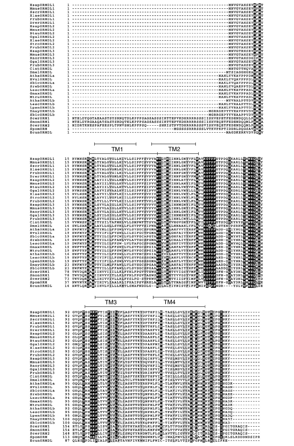
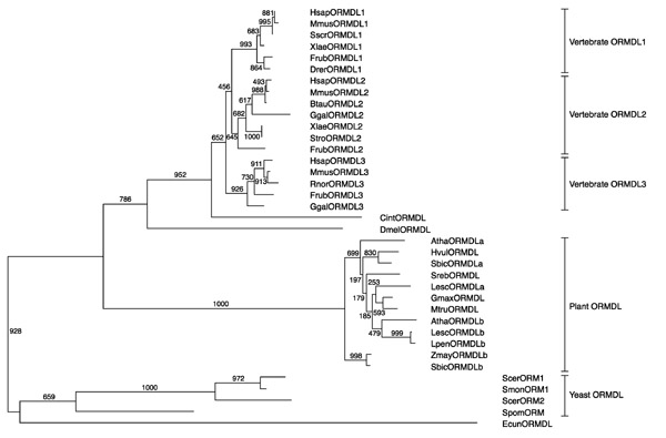
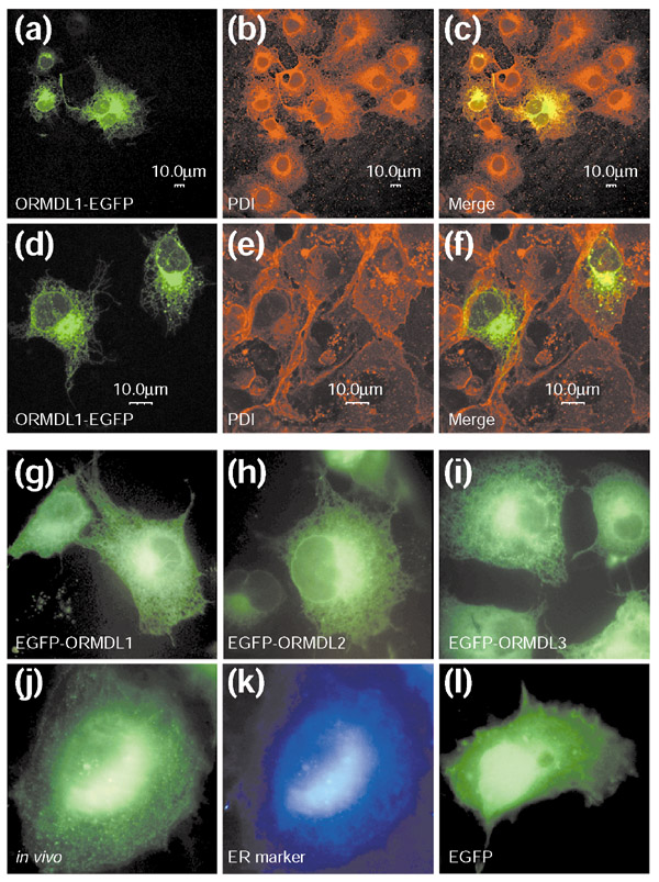
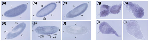
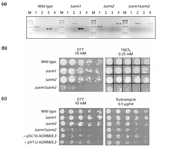
Results: While characterizing genes from the retinitis pigmentosa locus RP26 at 2q31-q33, we have identified a new gene, ORMDL1, that belongs to a novel gene family comprising three genes in humans (ORMDL1, ORMDL2 and ORMDL3), and homologs in yeast, microsporidia, plants, Drosophila, urochordates and vertebrates. The human genes are expressed ubiquitously in adult and fetal tissues. The Drosophila ORMDL homolog is also expressed throughout embryonic and larval stages, particularly in ectodermally derived tissues. The ORMDL genes encode transmembrane proteins anchored in the endoplasmic reticulum (ER). Double knockout of the two Saccharomyces cerevisiae homologs leads to decreased growth rate and greater sensitivity to tunicamycin and dithiothreitol. Yeast mutants can be rescued by human ORMDL homologs.
Conclusions: From protein sequence comparisons we have defined a novel gene family, not previously recognized because of the absence of a characterized functional signature. The sequence conservation of this family from yeast to vertebrates, the maintenance of duplicate copies in different lineages, the ubiquitous pattern of expression in human and Drosophila, the partial functional redundancy of the yeast homologs and phenotypic rescue by the human homologs, strongly support functional conservation. Subcellular localization and the response of yeast mutants to specific agents point to the involvement of ORMDL in protein folding in the ER.
Figures
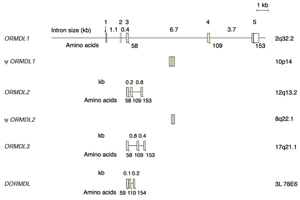
 ORMDL1, ORMDL2, and Drosophila ORMDL. Exon numbers of ORMDL1 are shown above the bars. Coding exons are shown in color and their sizes (in nucleotides) are: exon 3, 181 (orange); exon 4, 152 (yellow); exon 5, 133 (green). The intron sizes in kilobases are shown in-between the bars. The numbers below the bars denote the amino-acid positions for the exon-intron boundaries, and for the end of the ORF. The dotted appearance of the bar representing exon 2 of ORMDL1 indicates that it is alternatively spliced. The structure of the two human pseudogenes is also shown. The chromosomal location of each gene or pseudogene is indicated to the right.
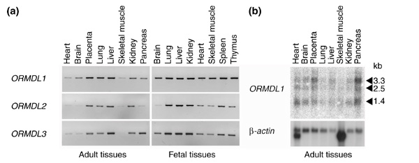
References
-
- International Human Genome Sequencing Consortium Initial sequencing and analysis of the human genome. Nature. 2001;409:860–921. - PubMed
-
- Adams MD, Celniker SE, Holt RA, Evans CA, Gocayne JD, Amanatides PG, Scherer SE, Li PW, Hoskins RA, Galle RF, et al. The genome sequence of Drosophila melanogaster. Science. 2000;287:2185–2195. - PubMed
-
- NCBI BLAST Server http://www.ncbi.nlm.nih.gov/BLAST/
Publication types
MeSH terms
Substances
Associated data
- Actions
- Actions
- Actions
- Actions
- Actions
- Actions
- Actions
- Actions
LinkOut - more resources
Full Text Sources
Other Literature Sources
Molecular Biology Databases
