Comparative Study
. 2002 Jul 1;21(13):3225-34.
doi: 10.1093/emboj/cdf332.

Crystal structure of the middle and C-terminal domains of the flagellar rotor protein FliG

Affiliations
Comparative Study

Crystal structure of the middle and C-terminal domains of the flagellar rotor protein FliG

Perry N Brown et al. EMBO J. .

Abstract

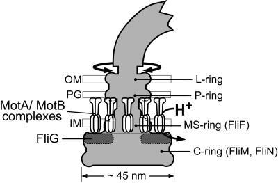
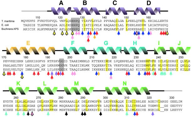
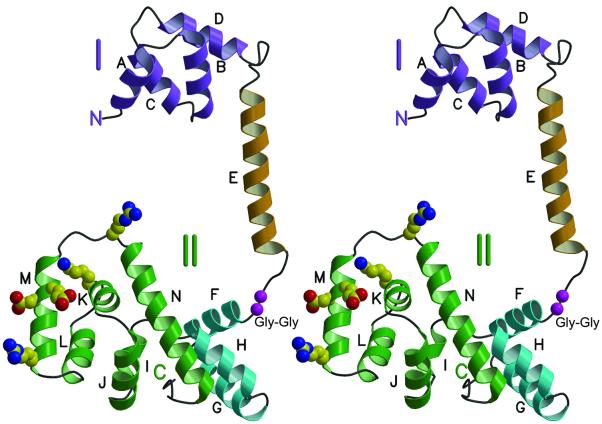
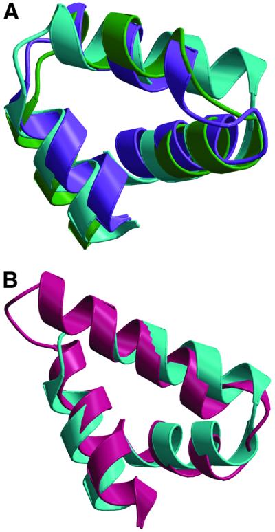
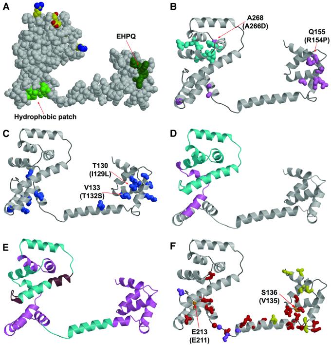
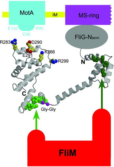
The FliG protein is essential for assembly, rotation and clockwise/counter-clockwise (CW/CCW) switching of the bacterial flagellum. About 25 copies of FliG are present in a large rotor-mounted assembly termed the 'switch complex', which also contains the proteins FliM and FliN. Mutational studies have identified the segments of FliG most crucial for flagellar assembly, rotation and switching. The structure of the C-terminal domain, which functions specifically in rotation, was reported previously. Here, we describe the crystal structure of a larger fragment of the FliG protein from Thermotoga maritima, which encompasses the middle and C-terminal parts of the protein (termed FliG-MC). The FliG-MC molecule consists of two compact globular domains, linked by an alpha-helix and an extended segment that contains a well-conserved Gly-Gly motif. Mutational studies indicate that FliM binds to both of the globular domains, and given the flexibility of the linking segment, FliM is likely to determine the relative orientation of the domains in the flagellum. We propose a model for the organization of FliG-MC molecules in the flagellum, and suggest that CW/CCW switching might occur by movement of the C-terminal domain relative to other parts of FliG, under the control of FliM.


Figures

None
Fig. 1. Schematic diagram of the E.coli flagellum, showing the approximate location of FliG. The shape of the rotor (shaded) is based on EM reconstructions of Francis et al. (1994). The stator is formed from multiple MotA–MotB complexes with probable composition (MotA)4(MotB)2 (Chun and Parkinson, 1988; DeMot and Vanderleyden, 1994; Sato and Homma, 2000; Braun and Blair, 2001). The placement of MotA–MotB complexes is based on freeze–fracture micrographs of Khan et al. (1988). IM, inner membrane; PG, peptidoglycan; OM, outer membrane.
None
Fig. 2. Alignment of FliG-MC sequences from T.maritima, E.coli and Buchnera APS. The initiating Met in the T.maritima protein studied here replaces a Leu in the native protein. Secondary structural elements seen in T.maritima FliG-MC are shown above. A thin line denotes disordered segments not included in the refined model. Residues that form the cores of domains I and II are highlighted in yellow. Conserved EHPQ and GG motifs discussed in the text are in gray boxes. Black arrows indicate charged residues important for motor rotation in E.coli (Lloyd and Blair, 1997). Colored arrows indicate positions of mutations giving various phenotypes. Turquoise, nonmotile but flagellate (Irikura et al., 1993; Lloyd et al., 1996). Pink, nonflagellate (in E.coli) (Lloyd et al., 1996; Zhou et al., 1998b). Blue, weakened binding to FliM in a yeast two-hybrid assay (Marykwas and Berg, 1996). Red, CW motor bias (Irikura et al., 1993; Lloyd and Blair, 1997). Yellow with black outline, CCW motor bias (Irikura et al., 1993). Orange, either CW or CCW motor bias depending on the substitution (Irikura et al., 1993). Purple, suppressors of mutations in the stator protein MotB (Garza et al., 1996). Most of the point mutations that gave nonflagellate phenotype in E.coli (pink arrows) gave an immotile but flagellate phenotype in Salmonella (Irikura et al., 1993); two exceptions that gave nonflagellate phenotype in both species are indicated by pink arrows with black outline. Sequences are from DDBJ/EMBL/GenBank.
None
Fig. 3. Stereoview ribbon representation of the FliG-MC structure. Helices are labeled A through N, and are colored according to domain. Purple, domain I. Gold, inter-domain helix. Green, the part of domain II whose structure was reported previously (Lloyd et al., 1999). Turquoise, the other part of domain II. Charged residues that interact with the stator and are important for motor rotation in E.coli are shown in standard atom colors. A well-conserved Gly–Gly sequence that should confer flexibility between the domains is shown in magenta.
None
Fig. 4. Superpositions of structural elements in FliG. (A) Overlay of helices B–D of FliG-MC (purple) with helices F–H (turquoise) and helices J–L (green). (B) Overlay of helices F–H of FliG-MC (turquoise) with residues 397–444 of β-catenin (protein data bank accession code 2BCT; Huber et al., 1997).
None
Fig. 5. Mapping of mutations and deletions to the structure. (A) Conserved surface features. Dark green, EHPQ motif in domain I. Light green, surface hydrophobic patch on domain II. Charged residues that interact with the stator are shown in standard atom colors. (B) Turquoise, mutations that eliminate motor rotation but allow flagellar assembly. Pink, mutations that disrupt flagellar assembly in E.coli. Mutations indicated by the labels (Q155 and A268) also disrupt flagellar assembly in Salmonella (compare with the legend to Figure 2; the mutational changes in Salmonella are given in parentheses). (C) Blue, mutations that weaken binding to FliM in the yeast two-hybrid assay (Marykwas and Berg, 1996). Two mutations that involved relatively minor changes in surface-exposed side-chains are indicated, with the mutational changes in the E.coli protein given in parentheses. These two mutations are adjacent to the EHPQ motif. (D) Turquoise, an 86-residue C-terminal segment that can be deleted while still allowing flagellar assembly and binding of FliG to FliM and FliN. Pink, a 19-residue segment whose further deletion prevents flagellar assembly and weakens binding to FliM and FliN (Tang et al., 1996). (E) Turquoise, segments of the protein in which 10-residue deletions prevent motor rotation but allow flagellar assembly. Pink, segments of the protein in which 10-residue deletions prevent flagellar assembly. Brown, segments of the protein in which 10-residue deletions allow both assembly and rotation. (F) Red, mutations giving CW motor bias (Irikura et al., 1993; Lloyd and Blair, 1997). Yellow, mutations giving CCW motor bias (Irikura et al., 1993). Orange, two positions that, depending on the substitution, give either CW or CCW motor bias; the residue numbers for T.maritima and Salmonella are shown (Salmonella in parentheses) (Irikura et al., 1993). Purple, mutations that suppress motility defects caused by mutations in the stator protein MotB (Garza et al., 1996).
None
Fig. 6. Proposed arrangement of FliG-MC relative to other proteins of the flagellum. The charge-bearing ridge on the C-terminal domain is oriented to allow interactions with conserved charged residues of MotA. FliM binds to both domains of FliG-MC, most likely through the EHPQ motif in domain I (dark green) and the surface hydrophobic patch on domain II (light green). (FliM–FliG binding interactions are indicated by the arrows.) The flexible Gly–Gly linker (magenta) allows relative movement of the domains. We propose that CW/CCW switching occurs by movement of the C-terminal domain of FliG relative to the rest of the protein, under the control of FliM (see the text).

References

    1. Aizawa S.-I., Dean,G.E., Jones,C.J., Macnab,R.M. and Yamaguchi,S. (1985) Purification and characterization of flagellar hook–basal body complex of Salmonella typhimurium. J. Bacteriol., 161, 836–849. - PMC - PubMed
    1. Asai J., Kojima,S., Kato,H., Nishioka,N., Kawagishi,I. and Homma,M. (1997) Putative channel components for the fast-rotating sodium-driven flagellar motor of a marine bacterium. J. Bacteriol., 179, 5104–5110. - PMC - PubMed
    1. Berg H.C. (2000) Constraints on models for the flagellar rotary motor. Phil. Trans. R. Soc. Lond. B Biol. Sci., 355, 491–501. - PMC - PubMed
    1. Berry R.M. and Armitage,J.P. (1999) The bacterial flagella motor. Adv. Microb. Physiol., 41, 291–337. - PubMed
    1. Blair D.F. (1995) How bacteria sense and swim. Annu. Rev. Microbiol., 49, 489–522. - PubMed

Publication types

Associated data