. 2002 Jul 1;21(13):3255-63.
doi: 10.1093/emboj/cdf351.

Cytoplasmic transport of Stat3 by receptor-mediated endocytosis

Affiliations

Cytoplasmic transport of Stat3 by receptor-mediated endocytosis

Andrea H Bild et al. EMBO J. .

Abstract

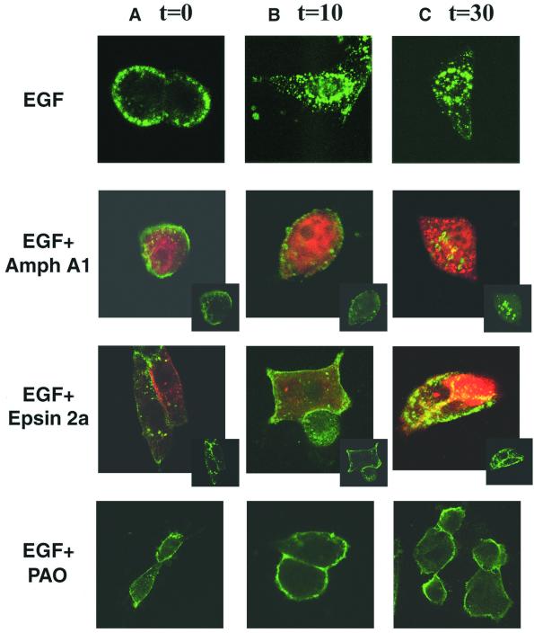
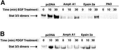
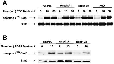
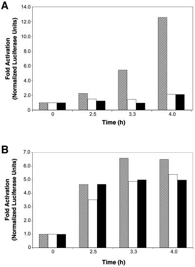
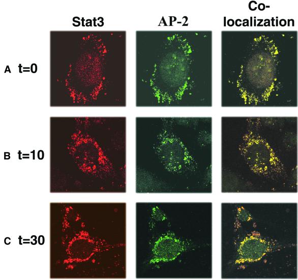
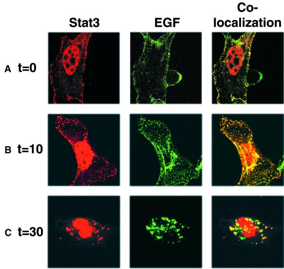
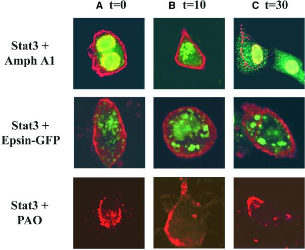
Signal transducer and activator of transcription (STAT) proteins are cytoplasmic transcription factors that translocate to the nucleus and regulate gene expression upon activation of cytokine or growth factor receptors. While this translocation event is essential for gene regulation by STATs, their mechanism of transport through the cytoplasm to the nucleus has remained elusive. We now report that cytoplasmic transport of Stat3 is an active process that requires receptor-mediated endocytosis. Stat3 co-localizes with endocytic vesicles in transit from the cell membrane to the perinuclear region in response to growth factor stimulation. Consistent with a role for receptor endocytosis in growth factor signaling, disruption of endocytosis with specific inhibitors blocks Stat3 nuclear translocation and Stat3-dependent gene regulation. These results indicate that receptor-mediated endocytosis may be a general mechanism of transport through the cytoplasm for a subset of cytoplasmic signaling proteins destined for the nucleus.


Figures

None
Fig. 1. Disruption of the endocytic machinery blocks EGFR internalization. Immunofluorescence analysis of NIH-3T3/EGFR cells was performed after transfection of expression vectors encoding HA-tagged Amph A1 or Epsin 2a, or treatment with 5 µM PAO. Antibodies to the HA tag of Amph A1 and Epsin 2a were used to detect localization of these proteins (red). Control (EGF alone) or treated cells (transfected or PAO treated) were incubated with 2 µg/ml Alexa Fluor EGF (green) for 45 min at 4°C to recruit ligand-bound receptor at the cell surface (A, time 0), and then warmed to 37°C for 10 min (B) and 30 min (C) to enable endocytosis. Localization of the EGF receptor–ligand complexes was analyzed using the LSM 510 program on a Zeiss confocal microscope. Insets show green fluorescence alone.
None
Fig. 2. Nuclear Stat3 DNA-binding activity is inhibited by blocking endocytosis. (A and B) DNA-binding activity of Stat3 complexes in either EGF-treated NIH-3T3/EGFR (A) or PDGF-BB-treated Balb/c-3T3 (B). Cells were transfected with empty vector control (pcDNA) or vectors encoding the endocytosis inhibitors Amph A1 or Epsin 2a, or treated with 5 µM PAO. Nuclear extracts were prepared following treatment with growth factors for the indicated times and were then subjected to EMSA analysis to detect activated Stat3 dimers bound to a 32P-labeled hSIE probe.
None
Fig. 3. Stat3 tyrosine phosphorylation is independent of nuclear translocation and receptor endocytosis. Western blot analyses were performed using antibodies to phosphoY705-Stat3 or total Stat3 protein to probe immunoprecipitates prepared with antibodies to total Stat3 protein from lysates of either EGF-treated NIH-3T3/EGFR (A) or PDGF-BB-treated Balb/c-3T3 (B) cells. Cells were treated with the endocytosis inhibitors followed by growth factor stimulation for the indicated times.
None
Fig. 4. Disruption of endocytosis with specific inhibitors blocks Stat3-dependent gene regulation. Balb/c-3T3 cells were transfected with control empty vector DNA (hatched) or expression vectors encoding the endocytosis inhibitors Amph A1 (open) or Epsin 2a (solid), the β-galactosidase vector as an internal control for transfection efficiency, and either the Stat3-specific luciferase reporter construct pLucTKS3 (A) or the Stat3-independent reporter construct pLucSRE (B). The transfected cells were harvested 48 h post-transfection following serum starvation overnight and treatment with 50 ng/ml PDGF for the times indicated. Results are shown as fold luciferase activities normalized to the β-gal internal control and are representative of three independent experiments.
None
Fig. 5. Stat3 localizes sequentially to endocytic vesicles at the cell membrane (A), in the cytosol (B) or at the perinuclear region (C) following PDGF treatment. (A–C) NIH-3T3 cells were treated with 50 ng/ml PDGF for 45 min at 4°C to recruit ligand-bound receptors at the cell surface, and then warmed to 37°C for the times indicated, to enable endocytosis. Staining with rabbit anti-Stat3 (red) or mouse anti-AP-2 (green) antibodies shows Stat3 in endosomes following growth factor stimulation. Images were collected using the LSM 510 program on a Zeiss confocal microscope.
None
Fig. 6. Stat3 co-localizes with Alexa Fluor EGF sequentially at the cell membrane (A), in the cytoplasm (B) or the perinuclear region (C) following growth factor stimulation. (A–C) NIH-3T3/EGFR cells overexpressing Stat3 were treated with 2 µg/ml Alexa Fluor EGF for 45 min at 4°C to recruit ligand-bound receptor at the cell surface, and then warmed to 37°C for the times indicated, to enable endocytosis. Immunofluorescent staining with rabbit anti-Stat3 antibodies was performed as in Figure 5. Images were collected using the LSM 510 program on a Zeiss confocal microscope.
None
Fig. 7. Inhibition of endocytosis blocks Stat3 translocation to the perinuclear region. Immunofluorescence analysis of NIH-3T3/EGFR cells was carried out after transfection of expression vectors encoding Stat3, Amph A1 or Epsin 2a–GFP. Antibodies to Stat3 or the HA tag of Amph A1 were used to detect localization of these proteins. Transfected cells were treated with 1 µg/ml EGF for 0 min (A), 10 min (B) and 30 min (C). Localization of Stat3 (red) and Amph A1 (green) or Epsin 2a–GFP (green) was analyzed using a Zeiss confocal microscope.

References

    1. Bowman T., Garcia,R., Turkson,J. and Jove,R. (2000) STATs in oncogenesis. Oncogene, 19, 2474–2488. - PubMed
    1. Bromberg J.F., Wrzeszczynska,M.H., Devgan,G., Zhao,Y., Pestell,R.G., Albanese,C. and Darnell,J.E.,Jr (1999) Stat3 as an oncogene. Cell, 98, 295–303. - PubMed
    1. Burke P., Schooler,K. and Wiley,H.S. (2001) Regulation of epidermal growth factor receptor signaling by endocytosis and intracellular trafficking. Mol. Biol. Cell, 12, 1897–1910. - PMC - PubMed
    1. Cahill M.A., Janknecht,R. and Nordheim,A. (1996) Signalling pathways: Jack of all cascades. Curr. Biol., 6, 16–19. - PubMed
    1. Ceresa B.P. and Schmid,S.L. (2000) Regulation of signal transduction by endocytosis. Curr. Opin. Cell Biol., 12, 204–210. - PubMed

Publication types

MeSH terms