. 2002 Jul 1;21(13):3307-16.
doi: 10.1093/emboj/cdf325.

Stress-inducible protein 1 is a cell surface ligand for cellular prion that triggers neuroprotection

Affiliations

Stress-inducible protein 1 is a cell surface ligand for cellular prion that triggers neuroprotection

Silvio M Zanata et al. EMBO J. .

Abstract

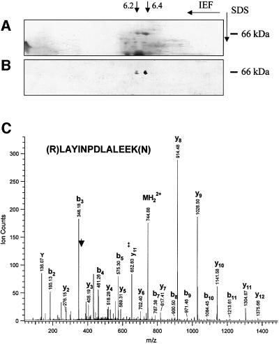
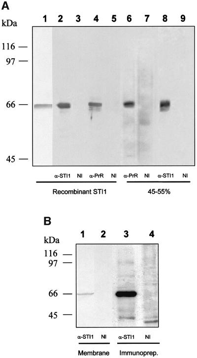
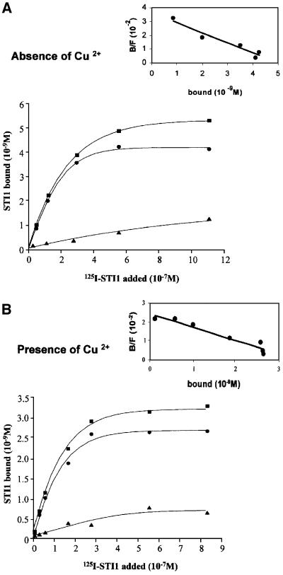
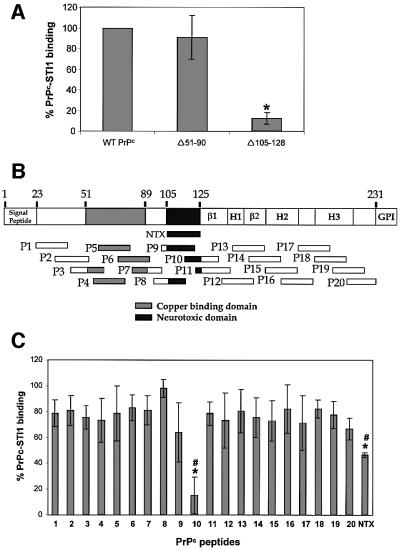
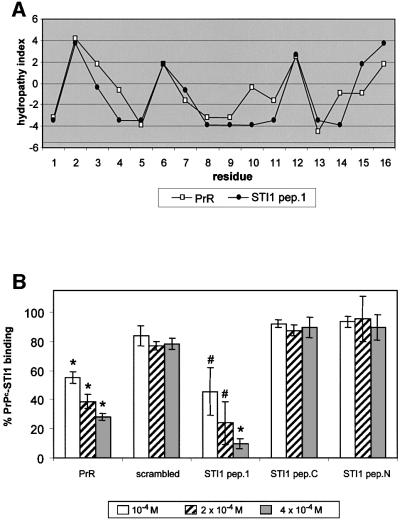
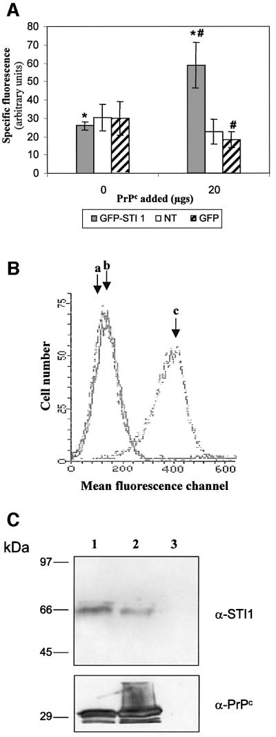
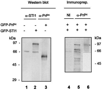
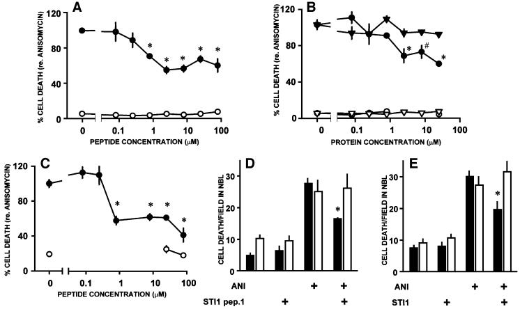
Prions are composed of an isoform of a normal sialoglycoprotein called PrP(c), whose physiological role has been under investigation, with focus on the screening for ligands. Our group described a membrane 66 kDa PrP(c)-binding protein with the aid of antibodies against a peptide deduced by complementary hydropathy. Using these antibodies in western blots from two-dimensional protein gels followed by sequencing the specific spot, we have now identified the molecule as stress-inducible protein 1 (STI1). We show that this protein is also found at the cell membrane besides the cytoplasm. Both proteins interact in a specific and high affinity manner with a K(d) of 10(-7) M. The interaction sites were mapped to amino acids 113-128 from PrP(c) and 230-245 from STI1. Cell surface binding and pull-down experiments showed that recombinant PrP(c) binds to cellular STI1, and co-immunoprecipitation assays strongly suggest that both proteins are associated in vivo. Moreover, PrP(c) interaction with either STI1 or with the peptide we found that represents the binding domain in STI1 induce neuroprotective signals that rescue cells from apoptosis.


Figures

None
Fig. 1. Two-dimensional gel analysis of the ammonium sulfate 45–55% saturation fraction from total brain extract. (A) Coomassie Blue-stained proteins. (B) Immunoblot of an identical gel reacted with mouse serum against the PrR peptide developed using peroxidase-labeled anti-mouse Ig. Two spots of 66 kDa and pIs of ∼6.2 and 6.4 are recognized specifically, and each was subjected to microsequencing analysis. (C) MS spectrum of a doubly charged tryptic peptide (MH22+ at m/z 744.9) from the spot with the higher pI. A series of fragment ions (b and y ions) were observed due to the breakage of peptide bonds during collision-induced dissociation. The peptide sequence is determined as LAYINPDLALEEK, identifying the protein as mouse stress-inducible protein 1 (accession No. 881485).
None
Fig. 2. STI1 is the 66 kDa protein located at the cell membrane recognized by serum against the PrR peptide. (A) Western blot assay of recombinant mSTI1 (lanes 2–5) or ammonium sulfate fractions at 45–55% saturation from brain extracts (lanes 6–9), done with rabbit serum against recombinant mST11 (α-STI1, lanes 2 and 8), serum against PrR peptide (α-PrR, lanes 4 and 6) or non-immune serum (NI, lanes 3, 5, 7 and 9). The recombinant STI1 protein stained with Ponceau is shown in lane 1. (B) Western blot assay from purified membrane fractions from brain extracts (lanes 1 and 2) done with rabbit serum against recombinant mST11 (α-STI1, lane 1) or non-immune serum (NI, lane 2). Cell surface proteins from N2a cells were biotinylated followed by extract preparation and immunoprecipitation with anti-STI1 antibody (α-STI1, lane 3) or non-immune serum (NI, lane 4). The immunoprecipitated material was developed using streptavidin– peroxidase.
None
Fig. 3. STI1 binds PrPc in a saturable and specific manner and independently of Cu2+ incorporation into the PrPc molecule. Representative curves of [125I]His6-STI1 binding to His6-PrPc refolded in the absence (A) or presence (B) of Cu2+. [125I]His6-STI1 was incubated with adsorbed His6-PrPc in the absence (total) or presence of unlabeled STI1 (non-specific). Non-specific binding (triangles) was subtracted from the total binding (squares) to yield His6-PrPc-specific binding to [125I]His6-STI1 (circles). Scatchard plots (inserts) gave Kds of 1.4 × 10–7 and 1.2 × 10–7 M for PrPc refolded in the absence or presence of Cu2+, respectively.
None
Fig. 4. Mapping the STI1 binding site in PrPc using deletion mutants and synthetic PrPc peptides. (A) Wild-type PrPc and deletion mutants Δ51–90 and Δ105–128 were incubated with [125I]His6-STI1. The binding between wild-type His6-PrPc and [125I]His6-STI1 was set to 100% (control). The results for each PrPc mutant were expressed as percentage binding compared with wild-type. *P <0.01 versus control, single mean Student’s t-test. (B) Twenty mouse PrPc peptides covering the PrPc (23–231) protein sequence were synthesized chemically as a 20mer with 10 overlapping residues. The scheme shows localization of the 20 peptides, the neurotoxic peptide (NTX) and the main PrPc domains: β1 and β2, β-sheet domains; H1, H2 and H3, α-helix domains; GPI, GPI anchor. (C) The synthetic peptides were pre-incubated with [125I]His6-STI1 followed by incubation with adsorbed His6-PrPc. Total binding between His6-PrPc and [125I]His6-STI1 was set to 100%. The results are expressed as the relative percentage of the binding produced by competition with each peptide. *P < 0.01 versus control, single mean Student’s t-test; #P < 0.01 NTX versus P10, Mann– Whitney test.
None
Fig. 5. Mapping PrPc binding at the STI1 molecule using the complementary hydropathy theory and synthetic peptides. (A) Hydropathy plot of STI1 pep.1 (amino acids 230–245) (filled circles) and the PrR peptide (open squares). (B) Competition of His6-PrPc–[125I]His6-STI1 binding by increasing amounts of the synthetic peptides PrR, scrambled peptide, STI1 pep.1, STI1 N-terminus peptide (STI1 pep.N) or STI1 C-terminus peptide (STI1 pep.C). Total binding between His6-PrPc and [125I]His6-STI1 was set to 100% (control). The results are expressed as the relative percentage of the binding produced by competition with each peptide. #P < 0.04 and *P < 0.01 versus control, single mean Student’s t-test.
None
Fig. 6. Cellular STI1 binds recombinant PrPc. (A) HEK 239T cells transfected with GFP–STI1 or GFP, or non-transfected (NT) were incubated in the absence or presence of 20 mg of His6-PrPc followed by incubation with mouse anti-PrPc or non-immune serum and anti-mouse R-phycoerythrin conjugate. Analyses were carried out using a Becton Dickinson FACScan Cytometer. The specific fluorescence intensity was determined by subtraction of the fluorescence obtained with non-immune serum from that produced with anti PrPc serum. *P <0.01, GFP–STI1 + 20 mg PrPc versus GFP–STI1 without PrPc and #P <0.03, GFP–STI1 + 20 mg PrPc versus GFP + 20 mg PrPc, Mann– Whitney test. (B) Primary fibroblast cultures from PrP0/0 animals were incubated in the absence (b) or presence (c) of His6-PrPc followed by incubation with mouse anti-PrPc (b and c) or non-immune serum (a). (C) Whole cells (lane 1) or cellular extracts (lane 2) from PrP0/0 mice fibroblasts were incubated with His6-PrPc. Whole cells were washed, lysed and the extracts incubated with Ni-NTA–agarose. Extracts from cells without His6-PrPc addition were also incubated with Ni-NTA–agarose (lane 3). The bound material was eluted off the beads and analyzed by western blot using anti-STI1 (α-STI1, upper panel) or anti-PrPc (α-PrPc, lower panel) serum.
None
Fig. 7. PrPc co-immunoprecipitates with STI1 located at the cell membrane. HEK 293T cells were transfected with GFP–PrPc and/or GFP–STI1 as indicated. Cell extracts were resolved by SDS–PAGE and western blots were done using anti-STI1 (lanes 1 and 2) or anti-PrPc (lane 3) antibodies. Cell surface proteins from transfected cells were biotinylated (lanes 4–6) and immunoprecipitated with anti-PrPc (lanes 5 and 6) or non-immune serum (lane 4). The reactions were developed using streptavidin–peroxidase (lanes 4 and 5) or anti-STI1 antibody followed by anti-rabbit IgG–peroxidase (lane 6). Lane 6 is shown only from 60 kDa upwards, because the antibody used for immunoprecipi tation reacts with the secondary antibody used to develop the western blot.
None
Fig. 8. Neuroprotective effects of STI1 and a PrPc-binding STI1 peptide. Cell death was counted within the neuroblastic layer of explants from the retinas of either neonatal rats or mice, maintained in vitro in various conditions. In (A–C), the rate of cell death was normalized with respect to the rate induced by anisomycin alone (100%). (A) Rates of cell death induced by anisomycin (1 µg/ml, filled circles) in retinal explants from neonatal rats, in the presence of various concentrations of the STI1 pep.1. Controls without anisomycin are shown with open circles. (B) Rates of cell death in retinal explants from neonatal rats, incubated with either recombinant STI1 (circles) or BSA (triangles), in either the presence (filled symbols) or absence (open symbols) of anisomycin. (C) Rates of cell death in explants from the retina of neonatal wild-type mice. Symbols as in (A). (D and E) Rates of cell death in retinal explants from neonatal wild-type (filled bars) or PrP0/0 (open bars) mice in various conditions. Note that both the STI1 pep.1 (25 µM, D) and the STI1 protein (8 µM, E) blocked anisomycin-induced cell death in wild-type, but not in PrP0/0 mice. All values are means ± SEM; *P < 0.01 and #P < 0.05 versus anisomycin alone, n ≥3 for each data point. Statistical significance is indicated only for the most relevant among the multiple comparisons tabulated in the Duncan’s test.

Similar articles

Cited by

References

    1. Aguzzi A. and Weissmann,C. (1997) Prion research: the next frontiers. Nature, 389, 795–798. - PubMed
    1. Ausubel F.M., Brent,K., Struhl,K., Kingston,R.E., Moore,D.D., Seidman,J.G. and Smith,J.A. (eds) (1993) Current Protocols in Molecular Biology. Wiley Interscience, New York.
    1. Beck K., Hunter,I. and Engel,J. (1990) Structure and function of laminin: anatomy of a multidomain glycoprotein. FASEB J., 4, 148–160. - PubMed
    1. Blatch G.L., Lassle,M., Takatori,T., Grandhi,T., Kundra,V. and Zetter,B.R. (1995) Molecular characterization of extendin: a protein localized in extending pseudopodia. Proc. Am. Assoc. Cancer Res., 36, 68.
    1. Blatch G.L., Lassle,M., Zetter,B.R. and Kundra,V. (1997) Isolation of a mouse cDNA encoding mSTI1, a stress-inducible protein containing the TPR motif. Gene, 194, 277–282. - PubMed

Publication types

MeSH terms