Comparative Study
. 2002 Jul 1;21(13):3358-69.
doi: 10.1093/emboj/cdf341.

Deconstructing PML-induced premature senescence

Affiliations
Comparative Study

Deconstructing PML-induced premature senescence

Oliver Bischof et al. EMBO J. .

Erratum in

Abstract

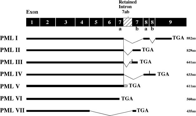
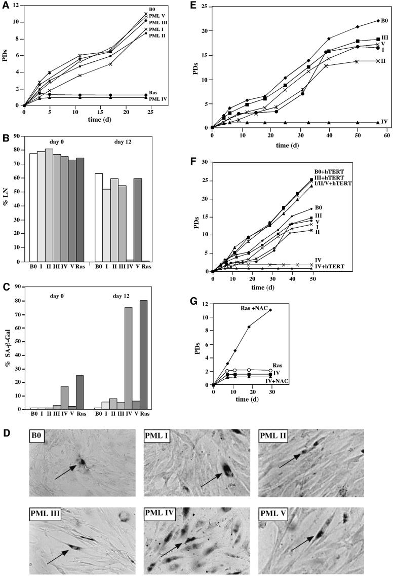
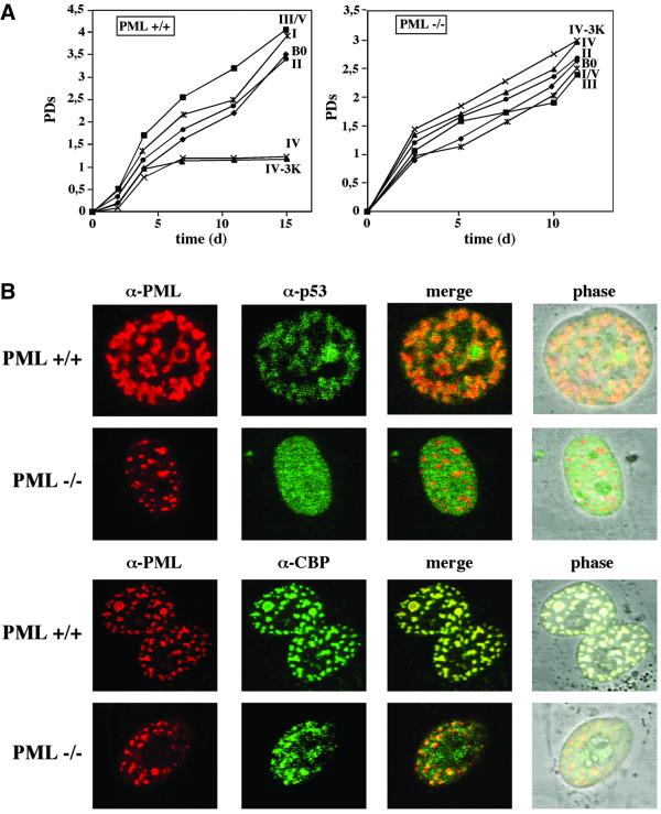
In this study, we investigated the subcellular and molecular mechanisms underlying promyelocytic leukemia (PML)-induced premature senescence. We demonstrate that intact PML nuclear bodies are not required for the induction of senescence. We have determined further that of seven known PML isoforms, only PML IV is capable of causing premature senescence, providing the first evidence for functional differences among these isoforms. Of interest is the fact that in contrast to PML(+/+) fibroblasts, PML(-/-) cells are resistant to PML IV-induced senescence. This suggests that although PML IV is necessary for this process to occur, it is not sufficient and requires other components for activity. Finally, we provide evidence that PML IV-induced senescence involves stabilization and activation of p53 through phosphorylation at Ser46 and acetylation at Lys382, and that it occurs independently of telomerase and differs from that elicited by oncogenic Ras. Taken together, our data assign a specific pro-senescent activity to an individual PML isoform that involves p53 activation and is independent from PML nuclear bodies.


Figures

None
Fig. 1. Schematic representation of the various PML isoforms. The classification of PML isoforms follows that of Jensen et al. (2001). The total number of amino acids (aa) for each isoform is given.
None
Fig. 2. Effect of PML isoforms on cell proliferation, DNA synthesis and induction of premature senescence. (A) Proliferation of normal human cells. WI38 normal human fibroblasts were infected with empty virus as a control (B0), oncogenic Ras- or PML-expressing retroviruses. Cells were selected for 4 days in 3 µg/ml puromycin and the number of population doublings (PDs) was determined over the indicated period of time. Day 0 is the first day after selection. PDs for each time point are the mean value of triplicates. (B and C) DNA synthesis and SA-β-Gal expression in normal human cells. WI38 cells were infected and selected as described in (A). After selection, [3H]thymidine was added for 3 days at the indicated time points (B), and cells were subsequently histochemically stained for SA-β-Gal expression (C) followed by autoradiography as described in Materials and methods. A minimum of 200 cells were counted to determine the percentages of positive SA-β-Gal expression and radiolabeled nuclei (%LN). A cell was only considered SA-β-Gal positive when it was not radiolabeled. PML isoforms are depicted in roman numbers. (D) Morphology and SA-β-Gal expression. Infected WI38 normal human fibroblasts were stained for SA-β-Gal as described and photographed under phase contrast optics. Arrows indicate examples of stained cells. Note the increased number of positive SA-β-Gal cells in PML IV-infected cells. (E) Life-span reduction by PML isoforms I, II, III and V. WI38 normal human fibroblasts were infected as described in (A) and serially passaged until the %LN dropped beyond 5% and the culture did not double for 3 weeks. (F) Telomerase and PML-induced growth arrest. Immortilized hTERT-WI38 fibroblasts were infected as described above and growth followed for the indicated period of time. The growth curves for I, II and V plus hTERT (I/II/V + hTERT) were superimposable. (G) WI38 normal human fibroblasts were infected with the indicated retroviruses as described in (A) and cultured in 2 mM NAC pre- and post-selection for the indicated period of time.
None
Fig. 3. Recruitment of p53 and CBP to PML NBs. (A and B) Representative images of WI38 normal human fibroblasts infected with B0 (control) or the respective PML-isoforms co-stained with either anti-PML (red) and anti-p53 (green) (A) or anti-CBP (green) (B), and analysed by laser scanning microscopy. The merged image of both stainings is shown in yellow. (C) Forced deposition of p53 into PML NBs. Representative images of WI38 normal human fibroblasts infected with a GFP fusion protein consisting of the minimal PML NB-targeting domain of SP100 and the p53 binding domain of MDM2 (SP–MDM2). SP–MDM2 was localized by direct GFP fluorescence (green) and p53 was visualized with an anti-p53 antibody (red). The merge appears in yellow. Note that SP–MDM2 colocalizes 100% with PML NBs (data not shown).
None
Fig. 4. Sumoylation and intact PML NBs are dispensable for premature senescence. WI38 normal human fibroblasts were infected with B0 (control), PML IV or the SUMO-deficient mutant PML IV-3K retroviruses as described in Figure 2, and growth curves (A), percentage of positive SA-β-Gal-stained cells and %LN (B) were determined. (C) Representative images of WI38 normal human fibroblasts infected with PML IV-3K, co-stained with either anti-PML (red) and anti-p53 (green) or anti-CBP (green), and analysed by laser scanning microscopy. (D–F) WI38 normal human fibroblasts were infected with IE1wt or IE1mt retroviruses and selected for 14 days in 400 µg/ml G418. Cells were then superinfected with either B0 (control), PML IV or oncogenic Ras retroviruses, and selected for 4 days in 3 µg/ml puromycin. (D) Representative images of WI38 normal human fibroblasts double-infected with either IE1wt+PML IV (a), IE1mt+PML IV (b) or IE1wt+Ras and IE1mt+Ras (c) retroviruses. Cells were co-stained either with anti-PML (red) and anti-p53 (green) or anti-CBP (green), and analysed by laser scanning microscopy. (E) Growth curves of WI38 fibroblasts co-infected with the indicated IE1mt+B0, IE1wt+B0, IE1wt+IV or IE1mt+IV, IE1wt+Ras or IE1mt+Ras retroviral constructs. IE1wt/mt+IV, cells transduced first with IE1wt or IE1mt followed by transduction with PML IV. (F) %LN and percentage of positive SA-β-Gal-stained cells from (E) at the indicated time points.
None
Fig. 5. PML IV stabilizes and activates p53. (A) WI38 normal human fibroblasts were infected with either B0 (control), PML III or PML IV retroviruses, as described in the legend to Figure 2. Cells were treated with 10 µg/ml cycloheximide for the indicated times when the PML IV-infected culture was terminally arrested, and whole-cell lysates were prepared and probed for p53. (B) WI38 normal human fibroblasts were infected with either B0 (control), PML I, III, IV, IE1wt or IE1wt+PML IV as described in the legend to Figure 2. Cell lysates were prepared when the PML IV-infected culture was terminally arrested, immunoprecipitated with goat anti-p53 antibody and analysed by western blot using anti-p53-acetylLys382 antibody. (C) Total lysates were prepared from WI38 normal human fibroblasts infected with B0 (control) or the indicated PML retroviruses as in Figure 2, and analysed by western blot using anti-p53-phosphoSer46 antibody and anti-QM antibody for normalization. (D) p53 transactivation activity in quiescent, senescent and PML-infected cells. WI38 fibroblasts were infected with the indicated PML and/or IE1wt retroviruses, and selected and transiently co-transfected with a pCMV-β-galactosidase normalization vector and the p21-Luc reporter when the PML IV-infected culture was completely growth arrested. Cells were assayed for β-galactosidase and luciferase reporters. Normalized p21 reporter activity is luciferase/ β-galactosidase activity, with the activity of p21-Luc in proliferating cells set at 1. Shown are the averages of three independent experiments. Within each experiment, transfections were performed in triplicate. Pre-senescent or senescent cells were cultured in 10% serum, in which case pre-senescent cells were growing exponentially, or incubated in 0.2% serum for 3 days, in which case pre senescent cells were quiescent.
None
Fig. 6. PML–/– MEFs are refractory to PML-induced premature senescence. (A) PML–/– MEFs or PML+/+ MEFs were infected with the respective PML retroviruses (in roman numbers) or empty vector control (B0), and growth curves were determined as described in the legend to Figure 2. (B) PML–/– MEFs do not recruit endogenous p53 to neoformed PML NBs. Representative images of PML–/– MEFs or PML+/+ MEFs infected with PML IV retroviruses and co-stained with either anti-PML (red) and anti-p53 (green) or anti-CBP (green), and analysed by laser scanning microscopy. (C) PML–/– MEFs show no increased acetylation of p53 at Lys382 upon PML overexpression. Immunoprecipitation of p53 from cells infected with the indicated retroviruses, and western blot analysis with anti-p53-acetylLys382 antibody as described in the legend to Figure 5.

References

    1. Alcalay M., Tomassoni,L., Colombo,E., Stoldt,S., Grignani,F., Fagioli,M., Szekely,L., Helin,K. and Pelicci,P.G. (1998) The promyelocytic leukemia gene product (PML) forms stable complexes with the retinoblastoma protein. Mol. Cell. Biol., 18, 1084–1093. - PMC - PubMed
    1. Appella E. and Anderson,C.W. (2001) Post-translational modifications and activation of p53 by genotoxic stresses. Eur. J. Biochem., 268, 2764–2772. - PubMed
    1. Bischof O., Kim,S.H., Irving,J., Beresten,S., Ellis,N.A. and Campisi,J. (2001) Regulation and localization of the Bloom syndrome protein in response to DNA damage. J. Cell Biol., 153, 367–380. - PMC - PubMed
    1. Bodnar A.G. et al. (1998) Extension of life-span by introduction of telomerase into normal human cells. Science, 279, 349–352. - PubMed
    1. Bringold F. and Serrano,M. (2000) Tumor suppressors and oncogenes in cellular senescence. Exp. Gerontol., 35, 317–329. - PubMed

Publication types

MeSH terms

Substances