. 2002 Jul 1;21(13):3402-13.
doi: 10.1093/emboj/cdf331.

Physiological and retinoid-induced proliferations of epidermis basal keratinocytes are differently controlled

Affiliations

Physiological and retinoid-induced proliferations of epidermis basal keratinocytes are differently controlled

Benoit Chapellier et al. EMBO J. .

Abstract

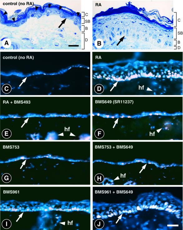
To investigate the roles of retinoic acid (RA) receptors (RARs) in the physiology of epidermis that does not express RAR beta, conditional spatio-temporally controlled somatic mutagenesis was used to selectively ablate RAR alpha in keratinocytes of RAR gamma-null mice. Keratinocyte proliferation was maintained in adult mouse epidermis lacking both RAR alpha and RAR gamma, as well as in RAR beta-null mice. All RAR-mediated signalling pathways are therefore dispensable in epidermis for homeostatic keratinocyte renewal. However, topical treatment of mouse skin with selective retinoids indicated that RXR/RAR gamma heterodimers, in which RXR transcriptional activity was subordinated to that of its RAR gamma partner, were required for retinoid-induced epidermal hyperplasia, whereas RXR homodimers and RXR/RAR alpha heterodimers were not involved. RA-induced keratinocyte proliferation was studied in mutant mice in which RXR alpha, RXR alpha and RAR alpha, RAR gamma, or RXR alpha and RAR gamma genes were specifically disrupted in either basal or suprabasal keratinocytes. We demonstrate that the topical retinoid signal is transduced by RXR alpha/RAR gamma heterodimers in suprabasal keratinocytes, which, in turn, stimulate proliferation of basal keratinocytes via a paracrine signal that may be heparin-binding EGF-like growth factor.


Figures

None
Fig. 1. Conditional mutagenesis of RARα in epidermis and homeostatic keratinocyte proliferation. (A) Schematic drawing of wild-type RARα locus (+), floxed L2 and excised (L) alleles. Black boxes stand for exons 7–9 (E7–9). Restriction sites and location of the 3′ probe are indicated. Sizes of restriction fragments are in kilobases (kb). H, HpaI; S, SacI. Arrowhead flags represent loxP sites. (B) Efficiency of Cre-ERT-mediated RARα gene disruption. Fourteen-week-old mice (genotypes as indicated) received TAM (5 days, 1 mg/day), and were treated again 2, 4 and 6 weeks later. RARα L2 and L alleles were identified on tail epidermis genomic DNA, before and 6 months after TAM injection. M: DNA ladder. (C) RARβ expression in skin samples. RNAse protection assay was performed on total RNA (20 µg) from control (RARαL2/L2) and mutant mice (RARγ–/– and RARαep–/–/RARγ–/–), 6 and 12 months after TAM administration. A tRNA sample and total RNAs from RA-treated F9 teratocarcinoma cells were used as controls. Histone H4 protection was included for quantitation of the RNA samples. (DF) Representative skin semi-thin sections, 12 months after TAM administration (genotypes as indicated). (GI) Representative skin sections labelled with BrdU (white colour), showing proliferation of basal keratinocytes 12 months after TAM administration (genotypes as indicated). Sections were counterstained with DAPI (blue colour). Arrows point to the dermal-epidermal junction. hf, hair follicles. Scale bar (in F and I): 50 µm.
None
Fig. 2. Histology and proliferative response of wild-type skin upon topical retinoid treatment. Dorsal skin was treated for four consecutive days with 40 nmol (in 400 µl acetone) of RA (B and D) or synthetic retinoids (E–J), as indicated. Controls (A and C) were treated with acetone vehicle. (A and B) Histology of control and RA-treated epidermis. Semi-thin sections (2-µm thick) were stained with toluidine blue. Arrowheads point to the spinous and granular keratinocytes in control epidermis. (CJ) Skin sections showing the proliferation marker Ki67 (white colour) and counterstained with DAPI (blue colour). Arrows point to the dermal-epidermal junction. RA, retinoic acid; BMS493, panRAR-selective antagonist; BMS649 (SR11237), panRXR-selective agonist; BMS753, RARα-selective agonist; BMS961, RARγ-selective agonist. B, basal layer; C, cornified layer; D, dermis; hf, hair follicle; SB, suprabasal layers (spinous and granular keratinocytes). Scale bar: 15 µm in (A) and (B); 25 µm in (C)–(I).
None
Fig. 3. Conditional mutagenesis of RXRα in epidermis, and RA-induced proliferative response. (A) Scheme of the experimental protocol. Intraperitoneal injection of TAM (1 mg) was performed from day 1 (D1) to day 4 (D4). A tail biopsy was made on day 11 to check for RXRα gene disruption. RA (40 nmol) was topically applied on skin for 4 days (D12–D15). On day 16 (D16), an injection of BrdU (50 mg/kg) was given 2 h before skin sampling. (B) Efficiency of RXRα gene disruption in mice bearing the K5-Cre-ERT transgene (as indicated). RXRα L2 and L alleles were identified on tail epidermis genomic DNA before and after TAM administration. M: DNA ladder. (C and D) Immunohistochemical detection of RXRα on skin sections from control (RXRαL2/L2) and mutant (RXRαep–/–) mice. (EJ) Representative skin sections labelled with BrdU (brown colour), showing epidermis thickness and basal keratinocyte proliferation in mutants (genotypes as indicated). Arrows point to the dermal–epidermal junction. hf, hair follicles. Scale bar: 25 µm in (C) and (D); 50 µm in (E)–(J).
None
Fig. 4. Conditional mutagenesis of RARγ and RA-induced proliferative response in mice lacking RARγ in suprabasal layers (RARγsb–/– mice). (A) Schematic drawing of RARγ wild-type (+), L3, partially excised L2Neo and fully excised L alleles. Sizes of NsiI fragments obtained for each allele are in kilobases (kb). Black boxes, exons 7–13 (E7–13); neo, neomycin gene; H, HpaI; N, NsiI. Arrowhead flags represent loxP sites. (B) Experimental protocol. Injection of TAM (1 mg) was performed from day 1 (D1) to day 3 (D3), then every second day for 10 days. A tail biopsy was made on day 9 to check for RARγ gene disruption. RA (40 nmol) was then applied topically on skin for 4 days (D10–D13). On day 14 (D14), the skin was sampled. (C) Efficiency of RARγ gene disruption in mice bearing the CMV-Cre-ERT transgene. RARγ wild-type (+), L3, L2Neo and L alleles were detected on tail epidermis and dermis genomic DNA before and after TAM administration. Note that the minute amount of RARγ excised L allele present in dermis most probably originates from contaminating epidermal keratinocytes (Li et al., 2000). M: DNA ladder. (DF) Representative skin sections showing RA-induced basal keratinocyte proliferation (white colour, Ki67 signal; blue colour, DAPI nuclear staining). RA-induced epidermal proliferation in RARγ–/– mice is illustrated in (F). (GI) Immunohistochemical detection of RARγ on back skin sections from control [(G), RARγsb+/–] and mutant [(H), RARγsb–/–] mice, after RA treatment. An RARγ–/– skin sample was used as negative control (I). Arrows point to the dermal–epidermal junction. hf, hair follicles. Scale bars (in F and I): 50 µm.
None
Fig. 5. Conditional mutagenesis of RXRα in epidermis suprabasal layers, and RA-induced proliferative response in RXRαsb–/– mice. (A) Efficiency of RXRα gene disruption in mice bearing the CMV-Cre-ERT transgene as compared with the K5-Cre-ERT transgene (as indicated). The experimental protocol is the same as in Figure 4B. RXRα wild-type (+), L2 and L alleles were detected on tail epidermis genomic DNA before and after TAM administration. M: DNA ladder. (B and C) Representative back skin sections labelled with BrdU (brown colour), showing RA-induced epidermal thickening and increased basal keratinocytes proliferation. (D and E) Immuno-histochemical detection of RXRα on skin sections from control [(D), RXRαsb+/–] and experimental [(E), RXRαsb–/–] mice, after RA treatment. Arrowheads indicate nuclei of suprabasal keratinocytes expressing RXRα. Arrows point to the dermal–epidermal junction. hf, hair follicles. Scale bar (in D): 50 µm.
None
Fig. 6. Expression of HB-EGF, CRABPII, RARβ2 and β-actin mRNA in whole back skin following topical application of retinoids. (A) Northern blot analysis of total RNA (25 µg) before (control) and after retinoid administration, as indicated. RA, retinoic acid; BMS493, panRAR antagonist; BMS961, RARγ agonist; BMS649 (SR11237), panRXR agonist. (B) Northern blot analysis of total RNA (25 µg) from control (RXRαL2/L2) and mutant mice (genotypes as indicated) before (–) and after (+) RA administration.

References

    1. Billoni N., Buan,B., Gautier,B., Gaillard,O., Mahé,Y.F. and Bernard,B.A. (2000) Thyroid hormone receptor β1 is expressed in the human hair follicle. Br. J. Dermatol., 142, 645–652. - PubMed
    1. Brocard J., Warot,X., Wendling,O., Messaddeq,N., Vonesch,J.L., Chambon,P. and Metzger,D. (1997) Spatio-temporally controlled site-specific somatic mutagenesis in the mouse. Proc. Natl Acad. Sci. USA, 94, 14559–14563. - PMC - PubMed
    1. Chambon P. (1996) A decade of molecular biology of retinoic acid receptors. FASEB J., 10, 940–954. - PubMed
    1. Chapellier B., Mark,M., Garnier,J.M., LeMeur,M., Chambon,P. and Ghyselinck,N.B. (2002a) A conditional floxed (loxP-flanked) allele for the Retinoic Acid Receptor alpha (RARα) gene. Genesis, 32, 87–89. - PubMed
    1. Chapellier B., Mark,M., Garnier,J.M., Dierich,A., Chambon,P. and Ghyselinck,N.B. (2002b) A conditional floxed (loxP-flanked) allele for the Retinoic Acid Receptor gamma (RARγ) gene. Genesis, 32, 95–98. - PubMed

Publication types

MeSH terms