Comparative Study
. 2002 Jul 1;21(13):3516-25.
doi: 10.1093/emboj/cdf333.

PA200, a nuclear proteasome activator involved in DNA repair

Affiliations
Comparative Study

PA200, a nuclear proteasome activator involved in DNA repair

Vicença Ustrell et al. EMBO J. .

Abstract

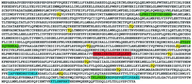
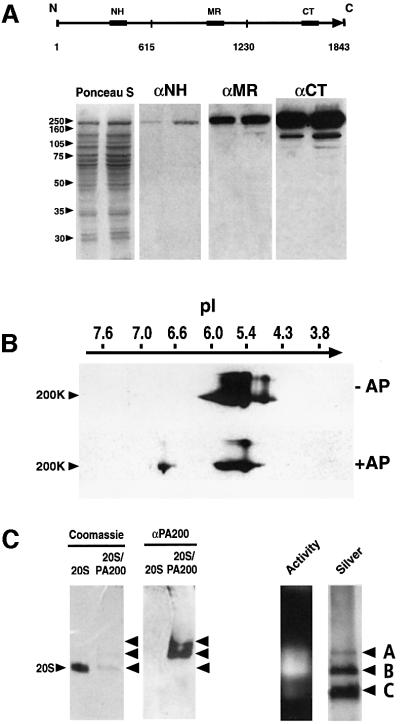
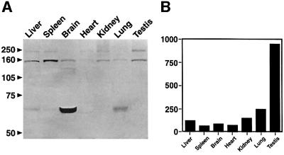
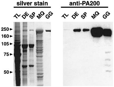
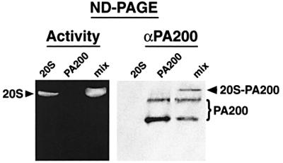
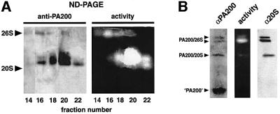
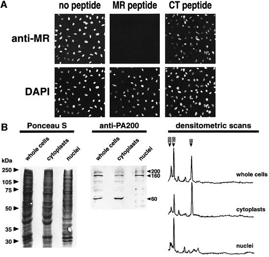
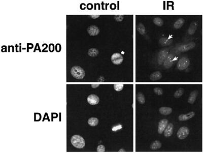
We have identified a novel 200 kDa nuclear protein that activates the proteasome. The protein, which we call PA200, has been purified to homogeneity from bovine testis and has been shown to activate proteasomal hydrolysis of peptides, but not proteins. Following gamma-irradiation of HeLa cells the uniform nuclear distribution of PA200 changes to a strikingly punctate pattern, a behavior characteristic of many DNA repair proteins. Homologs of PA200 are present in worms, plants and yeast. Others have shown that mutation of yeast PA200 results in hypersensitivity to bleomycin, and exposure of yeast to DNA damaging agents induces the PA200 message. Taken together, these findings implicate PA200 in DNA repair, possibly by recruiting proteasomes to double strand breaks.


Figures

None
Fig. 1. Amino acid sequence of human PA200. The amino acid sequence shown above was deduced from a human cDNA (KIAA0077) and analysis of human and mouse ESTs or genomes. The three peptides sequenced from rabbit reticulocyte PA200 are shown in blue, the three peptides (NH, MR, CT) used to generate antibodies are shown in green, the putative nuclear targeting sequence is highlighted in red, and yellow denotes 10 potential ATM phosphorylation sites (S/T-Q).
None
Fig. 2. Anti-peptide antibodies to PA200. (A) Western blot analysis of PA200 separated by SDS–PAGE. Samples (7 and 14 µg) of partially purified bovine testis PA200 were separated on 10% polyacrylamide gels and transferred to nitrocellulose. Pairs of lanes were then probed with a 1:1000 dilution of antiserum to each of the immunizing peptides, the location of which is shown schematically above. (B) Identification of PA200 isoforms by 2D PAGE. PA200 resolves into a series of isoelectric variants, at least one of which is phosphorylated based on alkaline phosphatase (AP) treatment. (C) Recognition by anti-PA200 of PA200–proteasome complexes separated on native gels. Proteasome– PA200 complexes and purified 20S proteasomes were separated in adjacent lanes of a 4.5% native acrylamide gel. A portion of the gel was stained directly with Coomassie Brilliant Blue (left) and an equivalent pair of lanes were transferred to nitrocellulose for western blotting with anti-PA200 (αPA200). Similar native gel analyses were performed in which proteasome activity was monitored by sLLVY-MCA over lay (activity) prior to fixation and silver staining of the same lane (far right). The species denoted A, B and C are thought to be PA200– proteasome–PA200 complexes, PA200–proteasome complexes and free 20S proteasomes, respectively.
None
Fig. 3. (A) Tissue distribution of PA200. Mouse organs were homogenized in 0.25%Triton X-100, 10 mM Tris pH 7.5, 1 mM DTT. Samples (20 µg) were resolved on 10% SDS–PAGE gels, transferred to nitrocellulose, and PA200 was detected by western blotting. Note that full-length PA200 is most abundant in testis, whereas the 160 kDa species is enriched in spleen and the 60 kDa immunoreactive protein is greatly enriched in brain. (B) PA200 mRNA levels in various human tissues and cell lines.The sequence of human PA200 was evaluated by a high-throughput gene expression profiling resource (Su et al., 2002; publicly available at http://expression.gnf.org).
None
Fig. 4. Purification of bovine testicular PA200. Fractions obtained during the purification of PA200 (see Supplementary data) were analyzed by SDS–PAGE and western blotting. Samples from testis lysate (TL), DEAE-pool (DE), a sizing pool (SP), a Mono Q pool (MQ) and the final glycerol gradient (GG) were separated on 10% SDS–PAGE gels and either silver stained (left) or transferred to nitrocellulose and probed with anti-PA200 (right).
None
Fig. 5. Proteasome activation by purified bovine PA200. Purified PA200 or 20S proteasomes were analyzed separately or mixed prior to electrophoresis on a native gel. Peptide overlay (left) demonstrated that PA200 activated the proteasome and western blotting (right) confirmed that some of the added PA200 bound the 20S proteasome (denoted 20S–PA200) in the right panel.
None
Fig. 6. Association of PA200 with the 26S proteasome. (A) Fractions obtained by DEAE chromatography of rabbit reticulocyte lysate enriched in 20S and 26S proteasomes were pooled and sedimented at 113 000 g for 22 h on a 25–45% glycerol gradient. The gradient was collected by bottom puncture, and samples of fractions 14, 16, 18, 20 and 22 were resolved on a 4.5% native polyacrylamide gels; the gels were overlaid with LLVY-MCA to detect 26S and 20S proteasomes (activity, right) prior to transfer to a nitrocellulose membrane for western blotting with anti-PA200 (left). Note that small amounts of anti-PA200 immunoreactivity coincides with 26S proteasome activity in fractions 14–20. (B) HeLa cells were extracted in 0.5% Triton X-100 and samples were analyzed in multiple lanes of a native acrylamide gel. Some lanes were overlaid with LLVY-MCA to localize 26S and 20S proteasomes (activity), while other lanes were transferred to nitrocellulose for western blotting with anti-PA200 (αPA200) or antibodies to 20S proteasome subunits (α20S). Note that as in (A), anti-PA200 immunoreactivity coincides with two forms of the 26S proteasome as well as with a 20S species. The component denoted ‘PA200’ (bottom left) represents the 60 kDa immunoreactive species.
None
Fig. 7. Intracellular distribution of PA200. (A) Indirect immunofluorescent HeLa cell stain. Cells growing on coverslips were fixed in 3.7% formaldehyde and stained with 1:300 anti-MR antiserum (see Figure 2) followed by FITC-goat anti-rabbit IgG. Note the excellent correspondence between staining by the DNA specific dye DAPI and FITC-fluorescence (left panels). Nuclear fluorescence was abolished when anti-MR was pre-incubated with 20 µg MR peptide prior to staining (center) but was not affected by preincubation with 20 µg CT peptide (right). (B) Distribution of PA200 in cytoplasts and nuclei prepared by cytochalasin-B-mediated enucleation. HeLa cells were enucleated as described in Materials and methods, and the distribution of PA200 was determined by western blotting of cytoplast and karyoplast proteins separated on 10% SDS–PAGE gels. The blot shown in the central panel and densitometry of it (right) clearly demonstrate that the 60 kDa immunoreactive species is confined to the cytoplasm, and that the 200 and 160 kDa species are located within nuclei.
None
Fig. 8. Intracellular distribution of PA200 following exposure of HeLa cells to ionizing radiation (IR). HeLa cells growing on coverslips were exposed to 50 Gy from a 137Cs source and fixed at 2, 6 or 22 h post-irradiation, then stained with anti-PA200 and DAPI. Note that the relatively uniform, albeit finely punctate, distribution of PA200 in control cells is distinctly different from the focal aggregates or foci of PA200 (white arrows) seen in HeLa nuclei 22 h after irradiation. Note also the even distribution of PA200 in the metaphase cell (control, asterisk).

References

    1. Anderson L., Henderson,C. and Adachi,Y. (2001) Phosphorylation and rapid relocalization of 53BP1 to nuclear foci upon DNA damage. Mol. Cell. Biol., 21, 1719–1729. - PMC - PubMed
    1. Baumeister W., Walz,J., Zhül,F. and Seemüller,E. (1998) The proteasome: paradigm of a self-compartmentalizing protease. Cell, 92, 367–380. - PubMed
    1. Bertolaet B.L., Clarke,D.J., Wolff,M., Watson,M.H., Henze,M., Divita,G. and Reed,S.I. (2001) UBA domains mediate protein–protein interactions between two DNA damage-inducible proteins. J. Mol. Biol., 313, 955–963. - PubMed
    1. Bischof O., Kim,S.-H., Irving,J., Beresten,S., Ellis,N.A. and Campisi,J. (2001) Regulation and localization of the bloom syndrome protein in reponse to DNA damage. J. Cell Biol., 153, 367–380. - PMC - PubMed
    1. Bochtler M., Ditzel,L., Groll,M., Hartmann,C. and Huber,R. (1999) The proteasome. Annu. Rev. Biophys. Biomol. Struct., 28, 295–317. - PubMed

Publication types

MeSH terms