. 2002 Jul 1;196(1):65-75.
doi: 10.1084/jem.20020201.

Chemokine requirements for B cell entry to lymph nodes and Peyer's patches

Affiliations

Chemokine requirements for B cell entry to lymph nodes and Peyer's patches

Takaharu Okada et al. J Exp Med. .

Abstract

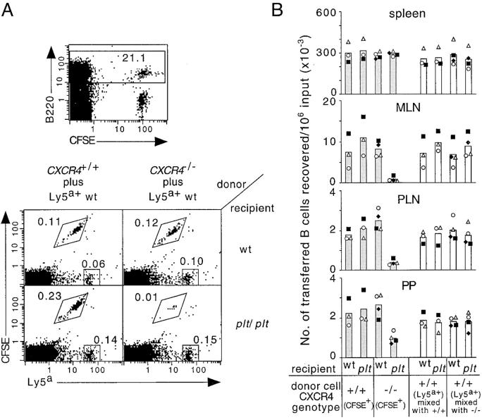
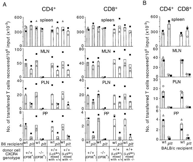
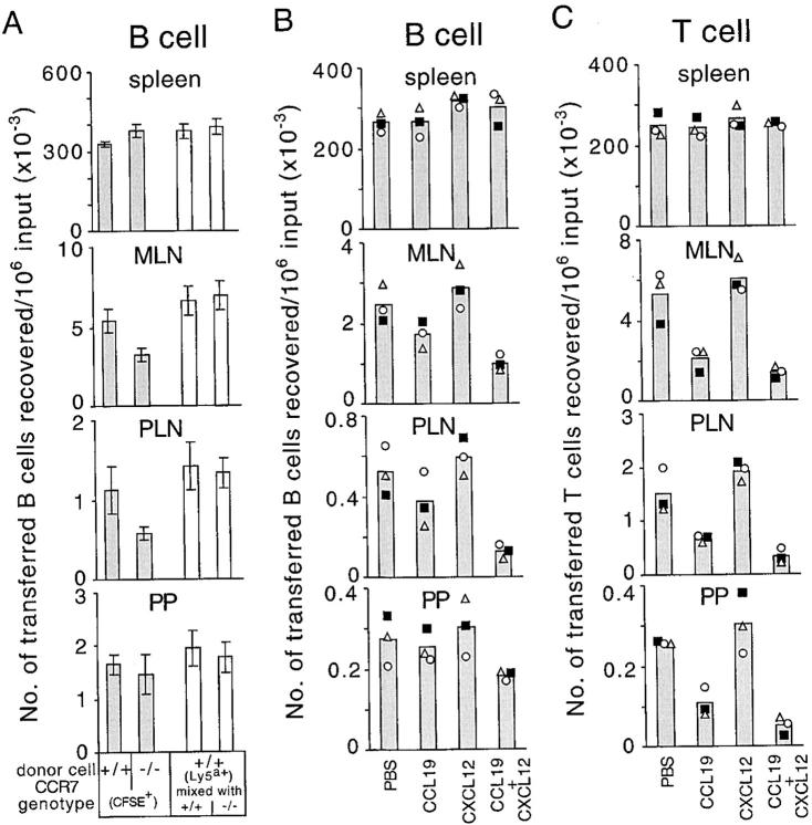
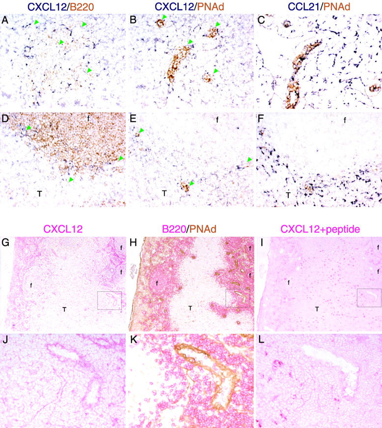
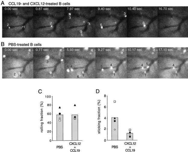
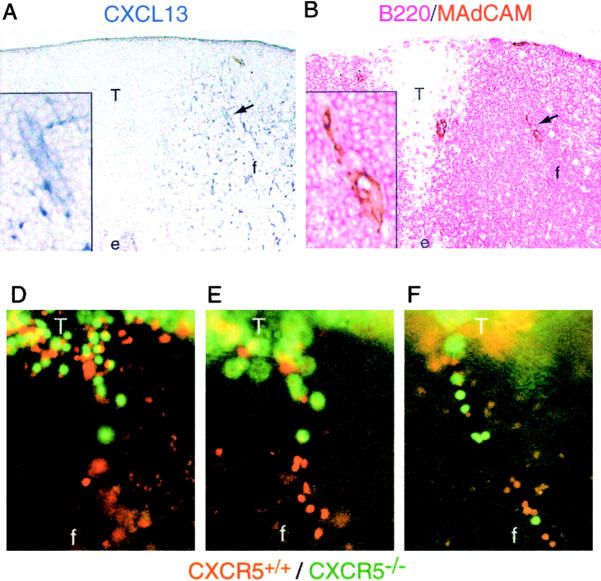
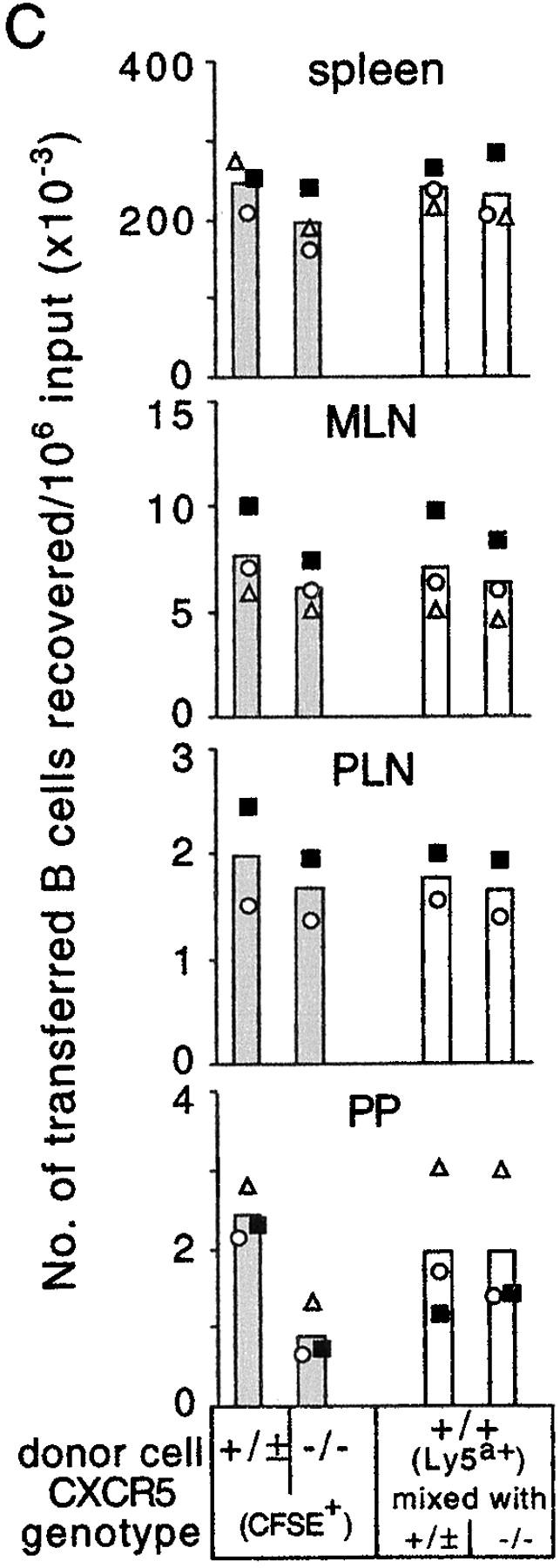
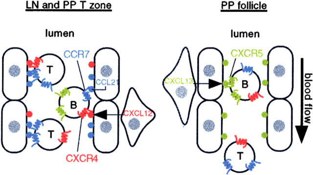
B cell entry to lymph nodes and Peyer's patches depends on chemokine receptor signaling, but the principal chemokine involved has not been defined. Here we show that the homing of CXCR4-/- B cells is suppressed in CCL19 (ELC)- and CCL21 (SLC)-deficient paucity of lymph node T cells mice, but not in wild-type mice. We also find that CXCR4 can contribute to T cell homing. Using intravital microscopy, we find that B cell adhesion to high endothelial venules (HEVs) is disrupted when CCR7 and CXCR4 are predesensitized. In Peyer's patches, B cell entry is dependent on CXCR5 in addition to CCR7/CXCR4. CXCL12 (SDF1) is displayed broadly on HEVs, whereas CXCL13 (BLC) is found selectively on Peyer's patch follicular HEVs. These findings establish the principal chemokine and chemokine receptor requirements for B cell entry to lymph nodes and Peyer's patches.


Figures

Figure 1.
Figure 1.
Defective homing of CXCR4-deficient B cells to lymph nodes and Peyer's patches of B6-plt/plt mice. (A) Representative flow cytometric analysis of transferred B cell accumulation in mesenteric lymph nodes of wild-type (wt) and B6-plt/plt recipients. CXCR4+/+ or CXCR4−/− splenocytes were CFSE labeled, mixed with wild-type control Ly5a+ splenocytes, and transferred into recipients for 90 min. Flow cytometric analysis of cells from a wild-type recipient of CXCR4+/+ plus Ly5a+ cells to show the separation of CFSE-labeled B and T cells, and the B220-gate (top). B220-gated cells plotted against CFSE fluorescence and Ly5a expression (bottom). Genotype of donor cell mixtures are indicated above the plots and recipient genotypes are indicated on the right of the plots. Gates demarcate the positions of CFSE+Ly5a− and CFSELy5a+ cells, and the numbers next to boxes indicate the percentage of total cells. (B) Enumeration of donor CXCR4+/+CFSE+Ly5a− B cells, CXCR4−/−CFSE+Ly5a− B cells (shaded columns) and CFSELy5a+ internal control B cells (open columns) recovered from spleen, mesenteric lymph nodes, inguinal and axillary lymph nodes, or four Peyer's patches of wild-type and B6-plt/plt recipients 90 min after transfer. Data are expressed as the number of CXCR4+/+, CXCR4−/−, or internal control B cells recovered in each recipient per 106 respective B cells transferred. Each symbol type represents an individual experiment. Columns show means of these individual data points. The number of transferred CXCR4−/− cells in B6-plt/plt recipient mesenteric lymph nodes, inguinal and axillary lymph nodes, and Peyer's patches was significantly lower than the number of CXCR4−/− cells in wt recipients (P < 0.01 in each case, paired t test), and significantly lower than the number of CXCR4+/+ and Ly5a+ wild-type cells in B6-plt/plt recipients (P < 0.005 in each case, Student's t test). wt, wild type; MLN, mesenteric lymph nodes; PLN, inguinal and axillary lymph nodes; PP, Peyer's patches.
Figure 2.
Figure 2.
CXCR4-dependent accumulation of T cells in lymph nodes and Peyer's patches of B6-plt/plt mice and T cell homing in BALB/c-plt/plt mice. (A) Enumeration of donor CXCR4+/+ or CXCR4−/− CD4 and CD8 T cells (shaded columns) and internal control CD4 and CD8 T cells (open columns) recovered from spleen, mesenteric lymph nodes, inguinal and axillary lymph nodes, or four Peyer's patches of wild-type and B6-plt/plt recipients 90 min after transfer. CXCR4+/+ or CXCR4−/− T cells were identified as CD3+CFSE+Ly5a− and internal control T cells as CD3+CFSELy5a+. These data are from three of the animals shown in Fig. 1. (B) Enumeration of donor CFSE+CD4+ or CFSE+CD8+ BALB/c T cells in BALB/c-plt/plt recipient tissues 90 min after transfer. Each symbol type represents an individual experiment and columns show means of these individual data points. The number of transferred wild-type CD4 and CD8 T cells in B6-plt/plt recipient mesenteric lymph nodes, inguinal and axillary lymph nodes, and Peyer's patches was significantly lower than the number that reached these tissues in wild-type recipients (P < 0.01 in each case, paired t test). The number of transferred CXCR4−/− CD4 and CD8 cells in B6-plt/plt recipient mesenteric lymph nodes, inguinal and axillary lymph nodes, and Peyer's patches was significantly reduced compared with the number of CXCR4+/+ and Ly5a+ wild-type cells reaching these organs in B6-plt/plt recipients (P < 0.03 in each case, Student's t test). MLN, mesenteric lymph nodes; PLN, inguinal and axillary lymph nodes; PP, Peyer's patches.
Figure 3.
Figure 3.
Suppression of B and T cell homing by desensitization of CCR7 and CXCR4. (A) Enumeration of donor CCR7+/+CFSE+Ly5a− B cells, CCR7−/−CFSE+Ly5a− B cells (shaded columns), and CFSELy5a+ internal control B cells (open columns) recovered from spleen, mesenteric lymph nodes, inguinal and axillary lymph nodes, or four Peyer's patches of wild-type recipients 90 min after transfer (mean ± SD; n = 7). The number of CCR7−/− B cells reaching mesenteric lymph nodes and inguinal and axillary lymph nodes was significantly reduced compared with CCR7+/+ cells (P < 0.05, paired t test). (B) Enumeration of donor CFSE+ B cells and (C) donor CFSE+ T cells, which had been incubated with PBS, 10 μg/ml CCL19 (ELC), 10 μg/ml CXCL12 (SDF1), or a combination of CCL19 and CXCL12 (10 μg/ml each) at 37°C for 45 min before transfer, recovered from spleen, mesenteric lymph nodes, inguinal lymph nodes, or three Peyer's patches (PP) of wild-type recipients 20 min after transfer. Each symbol type represents an individual experiment and columns show means of these individual data points. The number of CCL19 + CXCL12 pretreated B cells reaching mesenteric lymph nodes and inguinal lymph nodes was significantly reduced compared with PBS treated cells (P < 0.05, paired t test), whereas the effect in Peyer's patches did not reach statistical significance (P = 0.09). The number of CCL19 + CXCL12 pretreated T cells reaching inguinal and axillary lymph nodes was significantly reduced compared with CCL19 pretreated T cells (P < 0.05), whereas the effect in mesenteric lymph nodes and Peyer's patches was not statistically significant (P = 0.07 and 0.15, respectively). MLN, mesenteric lymph nodes; PLN, inguinal and axillary lymph nodes; PP, Peyer's patches.
Figure 4.
Figure 4.
Detection of CXCL12 (SDF1) on HEVs in lymphoid organs. (A–F) In situ hybridization analysis of CXCL12 and (C and F) CCL21 (SLC) mRNA expression (dark blue) in adjacent sections (A–C) of a mesenteric lymph node and (D–F) a Peyer's patch in combination with immunohistochemical staining (brown) for (A and D) B220 and (B, C, E, and F) PNAd to detect B cells and HEVs, respectively. Green arrowheads in A, B, D, and E indicate the positions of HEVs. Objective magnification, ×20. (G–L) Immunohistochemical analysis of CXCL12 protein distribution in lymph nodes. Adjacent sections of mesenteric lymph node were stained with anti-CXCL12 (G and J, red), anti-B220 (H and K, red), anti-PNAd (H and K, brown), and anti-CXCL12 antibody preincubated with CXCL12 blocking peptide (I and L, red). (J–L) High magnification images of the boxed areas in G–I. f, follicle; T, T zone. Objective magnification: G–I, ×5; J–L, ×20.
Figure 5.
Figure 5.
Combined desensitization of CXCR4 and CCR7 prevents B cell arrest on lymph node HEVs. (A) Representative time-lapse intravital micrographs of superficial inguinal lymph node HEVs, in which transferred 2-μM CFSE-labeled, chemokine-desensitized B cells flowed and rolled. Donor B cells had been incubated with a combination of CCL19 and CXCL12 (10 μg/ml each) before transfer. Transferred cells appearing in the vessel branches are numbered in their order of appearance. The time elapsed, starting from the first frame shown, is indicated in each image. B cells numbered 1–3 and 6 rolled on HEVs, but none of the cells became arrested. (B) Representative time-lapse intravital micrographs of the same superficial inguinal lymph node HEVs as in A, after secondary injection of 20-μM CFSE-labeled B cells, which had been incubated with PBS. B cells numbered 1 and 3–5 rolled on HEVs, and 1 and 4 were arrested on HEVs until the end of recording (∼5 min; see Videos 1 and 2 available at http://www.jem.org/cgi/content/full/ jem.20020201/DC1). (C and D) Fractions of B cells passing through HEVs that were recorded as (C) rolling or (D) arrested. B cells had been pretreated with PBS or with combined CCL19 and CXCL12 as in A and B. Arrested B cells were defined as cells that were adherent for greater than 1 min. Open symbols represent data from experiments in which chemokine-desensitized cells were injected first, and filled symbols represent data from experiments in which PBS-treated cells were injected first. Columns show means of the individual datasets. The number of CXCL12 + CCL19 pretreated cells that stuck was significantly reduced (P = 0.04, paired t test).
Figure 6.
Figure 6.
Involvement of CXCL13 (BLC) and CXCR5 in B cell homing to Peyer's patches. (A and B) Immunohistochemical analysis of CXCL13 distribution in Peyer's patches. Adjacent Peyer's patch sections were stained for (A) CXCL13 (blue), or B220 (red), and (B) MAdCAM (brown). A CXCL13+ HEV is indicated by the arrow, and higher magnification images of this vessel are shown in the insets. e, epithelium; f, follicle; T, T zone. Objective magnification, ×10 (insets, ×40). (C) Enumeration of donor CXCR5+/±CFSE+ B cells, CXCR5−/−CFSE+ B cells (shaded columns) and CFSELy5a+ internal control B cells (open columns) recovered from spleen, mesenteric lymph nodes, inguinal and axillary lymph nodes, or four Peyer's patches of wild-type recipients 90 min after transfer. In the transfer experiment shown by the filled squares, the CXCR5+/+ donor was CXCL13−/−. The number of transferred CXCR5−/− cells in Peyer's patches was significantly lower than the number of CXCR5+/+ cells (P < 0.001, paired t test). Similar findings were obtained in a separate experiment using CXCL13+/− recipients. MLN, mesenteric lymph nodes; PLN, inguinal and axillary lymph nodes; PP, Peyer's patches. (D–F) Whole mount microscopy of Peyer's patches from mice that had received CFSE-labeled CXCR5−/− and TRITC-labeled wild-type B cells 20 min earlier. Images in D and E are of the same field at different focal planes to provide detail of cells in the (D) T zone versus the (E) follicle. F shows a view of a vessel within another Peyer's patch. Objective magnification, ×20. The location of follicles and T cell areas was determined by orientating the Peyer's patch under low magnification (×5) and comparing the image with images obtained from similarly orientated Peyer's patches from animals that had received fluorescently labeled T cells. The data in D–F are representative of vessels observed in more than 10 Peyer's patches in three experiments. f, follicle; T, T cell.
Figure 6.
Figure 6.
Involvement of CXCL13 (BLC) and CXCR5 in B cell homing to Peyer's patches. (A and B) Immunohistochemical analysis of CXCL13 distribution in Peyer's patches. Adjacent Peyer's patch sections were stained for (A) CXCL13 (blue), or B220 (red), and (B) MAdCAM (brown). A CXCL13+ HEV is indicated by the arrow, and higher magnification images of this vessel are shown in the insets. e, epithelium; f, follicle; T, T zone. Objective magnification, ×10 (insets, ×40). (C) Enumeration of donor CXCR5+/±CFSE+ B cells, CXCR5−/−CFSE+ B cells (shaded columns) and CFSELy5a+ internal control B cells (open columns) recovered from spleen, mesenteric lymph nodes, inguinal and axillary lymph nodes, or four Peyer's patches of wild-type recipients 90 min after transfer. In the transfer experiment shown by the filled squares, the CXCR5+/+ donor was CXCL13−/−. The number of transferred CXCR5−/− cells in Peyer's patches was significantly lower than the number of CXCR5+/+ cells (P < 0.001, paired t test). Similar findings were obtained in a separate experiment using CXCL13+/− recipients. MLN, mesenteric lymph nodes; PLN, inguinal and axillary lymph nodes; PP, Peyer's patches. (D–F) Whole mount microscopy of Peyer's patches from mice that had received CFSE-labeled CXCR5−/− and TRITC-labeled wild-type B cells 20 min earlier. Images in D and E are of the same field at different focal planes to provide detail of cells in the (D) T zone versus the (E) follicle. F shows a view of a vessel within another Peyer's patch. Objective magnification, ×20. The location of follicles and T cell areas was determined by orientating the Peyer's patch under low magnification (×5) and comparing the image with images obtained from similarly orientated Peyer's patches from animals that had received fluorescently labeled T cells. The data in D–F are representative of vessels observed in more than 10 Peyer's patches in three experiments. f, follicle; T, T cell.
Figure 7.
Figure 7.
Principal chemokine requirements for B and T cell entry into lymph nodes and Peyer's patches. The diagram represents a lymph node or Peyer's patch T zone HEV on the left and a Peyer's patch follicular HEV on the right. CCL21 (SLC) is expressed by endothelial cells (oval shaped), whereas CXCL12 (SDF1) is expressed by stromal cells (triangular shaped) juxtaposed to HEVs. Both chemokines are presented on the lumenal side of HEVs. CCL19 (ELC) expressed by T zone stromal cells may also be presented on these HEVs (not shown). Large amounts of chemokine are also present in an abluminal location (not shown). CCR7 plays an indispensable role in triggering T cell adhesion, and CXCR4 can contribute partially to this process. B cells are triggered to adhere by either CCR7 or CXCR4, with CCR7 supporting approximately twofold more adhesion triggering events than CXCR4. Follicular areas of Peyer's patches contain HEVs of a smaller diameter than the T zone HEVs, and these vessels display CXCL13 (BLC), most likely derived from follicular stromal cells, although CXCL13 may also be expressed by the endothelial cells. B cells are triggered to adhere to these vessels by CXCL13/CXCR5. CXCR5 supports ∼50% of the chemokine triggering requirement for B cell homing to Peyer's patches. Most T cells do not express CXCR5 and are not triggered to adhere to HEVs in follicles, passing through to T zone HEVs where they may be arrested. T, T cells; B, B cells.

References

    1. Springer, T.A. 1994. Traffic signals for lymphocyte recirculation and leukocyte emigration: the multistep paradigm. Cell. 76:301–314. - PubMed
    1. Butcher, E.C., and L.J. Picker. 1996. Lymphocyte homing and homeostasis. Science. 272:60–66. - PubMed
    1. Spangrude, G.J., B.A. Braaten, and R.A. Daynes. 1984. Molecular mechanisms of lymphocyte extravasation I. Studies of two selective inhibitors of lymphocyte recirculation. J. Immunol. 132:354–362. - PubMed
    1. Bargatze, R.F., and E.C. Butcher. 1993. Rapid G protein regulated activation event involved in lymphocyte binding to high endothelial venules. J. Exp. Med. 178:367–372. - PMC - PubMed
    1. Cyster, J.G., and C.C. Goodnow. 1995. Pertussis toxin inhibits migration of B and T lymphocytes into splenic white pulp cords. J. Exp. Med. 182:581–586. - PMC - PubMed

Publication types

MeSH terms