. 2002 Jul 1;196(1):77-85.
doi: 10.1084/jem.20020439.

Critical roles for interleukin 1 and tumor necrosis factor alpha in antibody-induced arthritis

Affiliations

Critical roles for interleukin 1 and tumor necrosis factor alpha in antibody-induced arthritis

Hong Ji et al. J Exp Med. .

Abstract

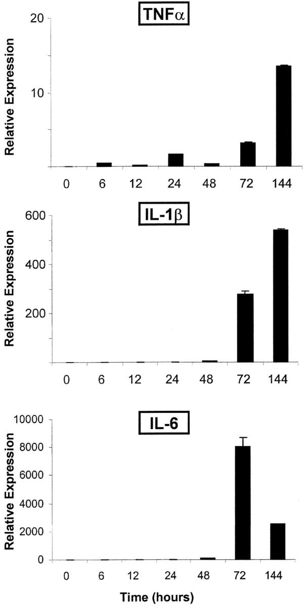
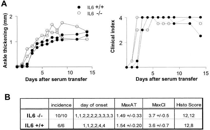
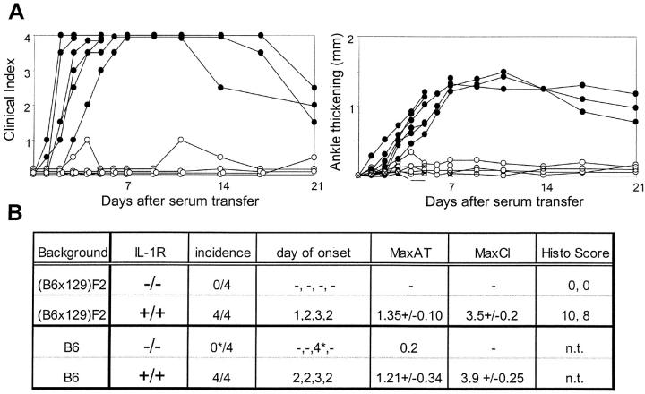
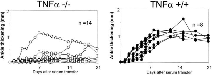
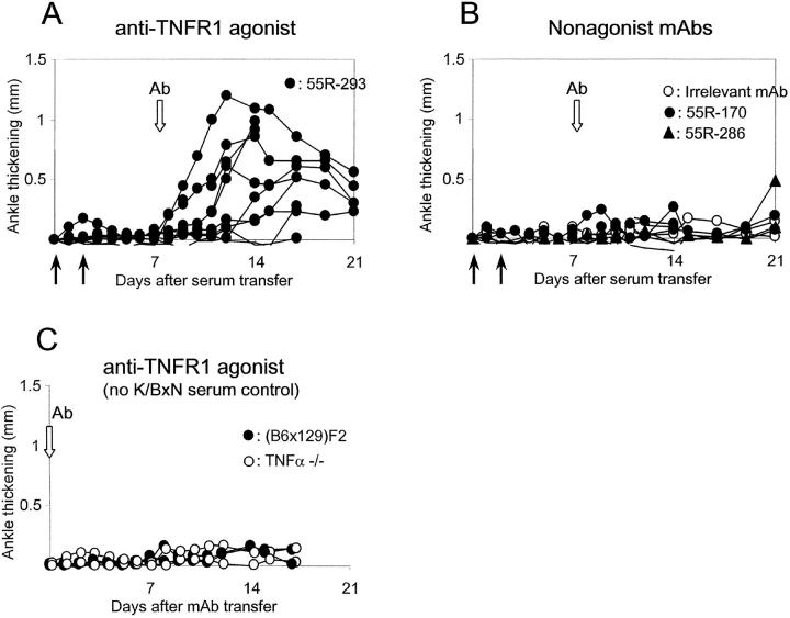
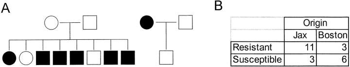
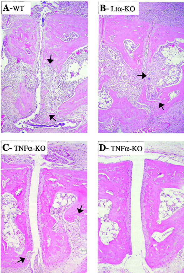
In spontaneous inflammatory arthritis of K/BxN T cell receptor transgenic mice, the effector phase of the disease is provoked by binding of immunoglobulins (Igs) to joint surfaces. Inflammatory cytokines are known to be involved in human inflammatory arthritis, in particular rheumatoid arthritis, although, overall, the pathogenetic mechanisms of the human affliction remain unclear. To explore the analogy between the K/BxN model and human patients, we assessed the role and relative importance of inflammatory cytokines in K/BxN joint inflammation by transferring arthritogenic serum into a panel of genetically deficient recipients. Interleukin (IL)-1 proved absolutely necessary. Tumor necrosis factor (TNF)-alpha was also required, although seemingly less critically than IL-1, because a proportion of TNF-alpha-deficient mice developed robust disease. There was no evidence for an important role for IL-6. Bone destruction and reconstruction were also examined. We found that all mice with strong inflammation exhibited the bone erosion and reconstruction phenomena typical of K/BxN arthritis, with no evidence of any particular requirement for TNFalpha for bone destruction. The variability in the requirement for TNF-alpha, reminiscent of that observed in treated rheumatoid arthritis patients, did not appear genetically programmed but related instead to subtle environmental changes.


Figures

Figure 1.
Figure 1.
Kinetic of inflammatory cytokine expression. Arthritis was induced by injection of K/BxN serum into naive C57Bl/6 mice, and RNA was prepared from ankle joint cavities at various points thereafter. mRNA encoding inflammatory cytokines were quantitated by real-time PCR using cyclophilin mRNA as an internal standard. The results are presented as relative expression of the individual cytokine mRNAs standardized against a reference sample of total spleen RNA from a normal mouse. This is a representative experiment (of three), with two mice pooled for each point.
Figure 2.
Figure 2.
No requirement for IL-6 in arthritis induced by K/BxN serum transfer. IL6-deficient and control mice (matched for gender/age and genetic background) were injected with 150 μl serum from arthritic K/BxN animals on days 0 and 2. Arthritis was evaluated by measuring clinical index and ankle thickening (Materials and Methods). (A) Data from a representative experiment, with each curve representing an individual mouse. (B) Tabulation of the results for 10 knockout mice and age/gender matched controls; MaxAT, maximum increase in ankle thickness in millimeters. MaxCI, maximum clinical index, sum of scores on each limb (for each limb: 0, no disease; 0.5, mild swelling of paw or of just a few digits; 1, clear joint inflammation in ankle or wrists); maximum score = 4. The histological score sums scores from knee, ankle, and tarsal joints (1, minimum synovial hyperplasia; 2, limited inflammatory infiltration; 3, massive infiltration; 4, massive infiltration with cartilage and bone destruction); maximum score = 12.
Figure 3.
Figure 3.
Essential role of IL-1 ILIR–deficient and control mice (matched for gender/age and genetic background) were injected with 150 μl serum from arthritic K/BxN animals on days 0 and 2. Arthritis was evaluated by measuring clinical index and ankle thickening as in Fig. 2. (A) Data from a representative experiment in B6 recipients, with each curve representing an individual mouse. (B) Tabulation of the results for eight knockout mice and age/gender matched controls on either the standard (B6 × 129)F2 background or on an inbred B6 background. Scoring as described for Fig. 2; the star denotes a transient inflammation in the digits of one mouse.
Figure 4.
Figure 4.
Variability of arthritis in TNFα-deficient mice. TNFα-deficient (left) and control mice (right; matched for gender/age and genetic background) were injected with 150 μl serum from arthritic K/BxN animals on days 0 and 2. Arthritis was evaluated by measuring ankle thickening as in Fig. 2. The data are pooled from six different experiments. All mice originated from the Jackson Laboratory.
Figure 5.
Figure 5.
Triggering the TNF receptor complements TNFα deficiency. TNFα-deficient mice were injected with 150 μl serum from arthritic K/BxN animals on days 0 and 2. Animals not presenting any signs of disease by day 7 were injected at days 7, 11, and 15 with anti-TNFR1 mAb 55R-293, which has significant agonist activity (A) or with control mAbs (B). These controls included anti-TNFR1 reagents devoid of agonist activity or an irrelevant mAb. (C) Anti-TNFR mAbs were injected without K/BxN serum. Arthritis was evaluated by measuring ankle thickening as above. The data are pooled from four different experiments. All mice originated from the Jackson Laboratory.
Figure 6.
Figure 6.
Environmental, not genetic, influences on TNF-independent arthritis. (A) TNFα-deficient mice from the Jackson Laboratory were tested by transfer of K/BxN serum, and animals of different phenotypes were crossed (white symbols, resistant mice; black symbols, susceptible mice, where resistance and susceptibility are defined as the presence of clear arthritis [grade > 1]) in the first 10 d after serum transfer. Their progeny was similarly tested when 4–5 wk old. (B) A compilation of results of challenge of TNFα-deficient mice with K/BxN serum, either from mice purchased from the Jackson Laboratory or bred in our Boston colony; χ2, P = 0.003.
Figure 7.
Figure 7.
Bone destruction in control and KO mice. Bone erosion was assessed in hematoxylin and eosin–stained ankle sections from TNFα- and LTα-deficient mice and control mice after transfer of K/BxN serum. (A) Control mouse, full inflammation and areas of focal bone destruction (arrows). (B) Arthritic LTα-deficient mouse with inflammation and focal bone erosion. (C) TNFα-deficient mouse with clinical manifestations, showing inflammation and focal bone erosion. (D) TNFα-deficient mouse with no clinically detectable symptoms, showing minimal inflammation (matched with A).

References

    1. Feldmann, M., and R.N. Maini. 2001. Anti-TNFα therapy of rheumatoid arthritis: what have we learned? Annu. Rev. Immunol. 19:163–196. - PubMed
    1. Aarvak, T., and J.B. Natvig. 2001. Cell-cell interactions in synovitis: antigen presenting cells and T cell interaction in rheumatoid arthritis. Arthritis Res. 3:13–17. - PMC - PubMed
    1. Firestein, G.S., and N.J. Zvaifler. 1990. How important are T cells in chronic rheumatoid synovitis? Arthritis Rheum. 33:768–773. - PubMed
    1. van den Berg, W.B. 2001. Anti-cytokine therapy in chronic destructive arthritis. Arthritis Res. 3:18–26. - PMC - PubMed
    1. Kouskoff, V., A.-S. Korganow, V. Duchatelle, C. Degott, C. Benoist, and D. Mathis. 1996. Organ-specific disease provoked by systemic autoreactivity. Cell. 87:811–822. - PubMed

Publication types

MeSH terms