. 2002 Jul 1;196(1):119-27.
doi: 10.1084/jem.20020092.

A critical role for natural killer T cells in immunosurveillance of methylcholanthrene-induced sarcomas

Affiliations

A critical role for natural killer T cells in immunosurveillance of methylcholanthrene-induced sarcomas

Nadine Y Crowe et al. J Exp Med. .

Abstract

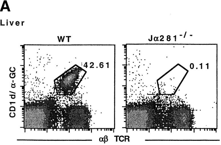
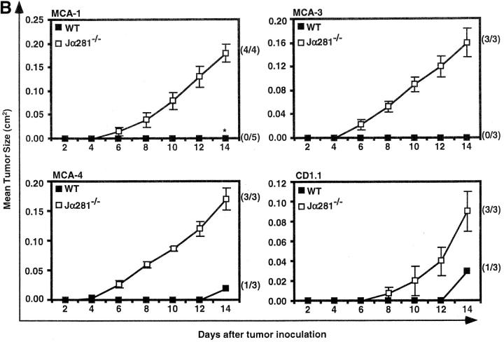
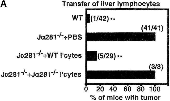
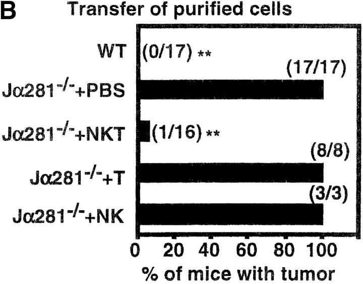
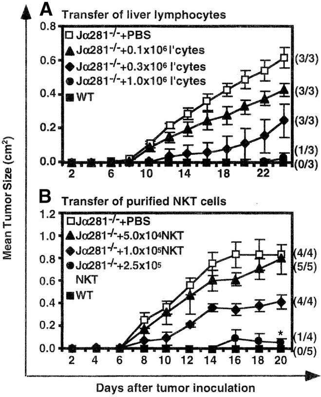
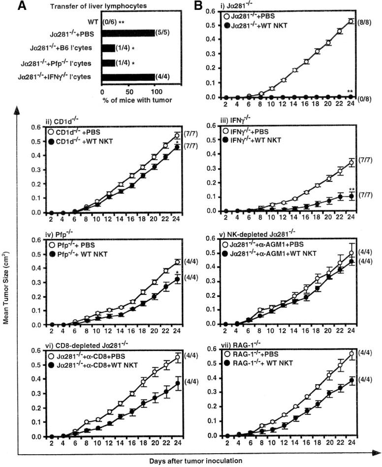
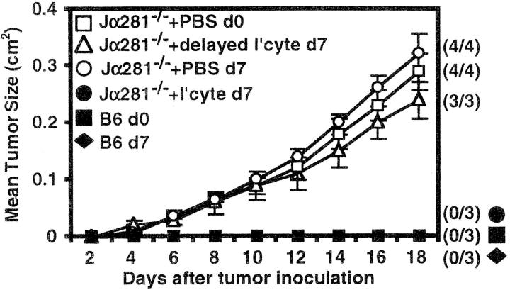
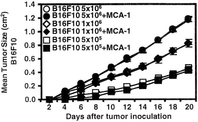
Natural killer (NK) T cells initiate potent antitumor responses when stimulated by exogenous factors such as interleukin (IL)-12 or alpha-galactosylceramide (alpha-GalCer), however, it is not clear whether this reflects a physiological role for these cells in tumor immunity. Through adoptive transfer of NK T cells from wild-type to NK T cell-deficient (T cell receptor [TCR] Jalpha281-/-) mice, we demonstrate a critical role for NK T cells in immunosurveillance of methylcholanthrene (MCA)-induced fibrosarcomas, in the absence of exogenous stimulatory factors. Using the same approach with gene-targeted and/or antibody-depleted donor or recipient mice, we have shown that this effect depends on CD1d recognition and requires the additional involvement of both NK and CD8+ T cells. Interferon-gamma production by both NK T cells and downstream, non-NK T cells, is essential for protection, and perforin production by effector cells, but not NK T cells, is also critical. The protective mechanisms in this more physiologically relevant system are distinct from those associated with alpha-GalCer-induced, NK T cell-mediated, tumor rejection. This study demonstrates that, in addition to their importance in tumor immunotherapy induced by IL-12 or alpha-GalCer, NK T cells can play a critical role in tumor immunosurveillance, at least against MCA-induced sarcomas, in the absence of exogenous stimulation.


Figures

Figure 1.
Figure 1.
NK T cell–deficient (Jα281−/−) mice are more susceptible to growth of MCA-induced sarcoma lines. (A) Liver lymphocytes were isolated from WT and Jα281−/− mice and labeled with CD1/α-GalCer tetramer and mAb specific for αβ TCR to identify NK T cells. (B) Groups of WT and Jα281−/− mice were injected subcutaneously (hind flank) with 105 MCA-1, -3, -4 or CD1.1 sarcoma cell lines. Results were recorded every other day as the mean tumor size (in cm2) ± SEM. Three mice per group were used. Significant difference from the PBS-treated Jα281−/− group was determined using a Mann-Whitney U test when n ≥ 4. *P ≤ 0.05.
Figure 1.
Figure 1.
NK T cell–deficient (Jα281−/−) mice are more susceptible to growth of MCA-induced sarcoma lines. (A) Liver lymphocytes were isolated from WT and Jα281−/− mice and labeled with CD1/α-GalCer tetramer and mAb specific for αβ TCR to identify NK T cells. (B) Groups of WT and Jα281−/− mice were injected subcutaneously (hind flank) with 105 MCA-1, -3, -4 or CD1.1 sarcoma cell lines. Results were recorded every other day as the mean tumor size (in cm2) ± SEM. Three mice per group were used. Significant difference from the PBS-treated Jα281−/− group was determined using a Mann-Whitney U test when n ≥ 4. *P ≤ 0.05.
Figure 2.
Figure 2.
Adoptive transfer of NK T cells protects Jα281−/− mice against MCA-1 tumor growth. Groups of WT and Jα281−/− mice were inoculated subcutaneously (hind flank) with 105 MCA-1 cells. (A) Subgroups of Jα281−/− mice then received between 1 and 3 × 10⁶ WT or 3 × 10⁶ Jα281−/− liver lymphocytes, 200 μL 2% NMS.PBS, or (B) 2.5 × 10⁵ purified NK1.1+αβ TCR+ (NK T), NK1.1+αβ TCR (NK), NK1.1 αβ TCR+ (T) cells, or 200 μL 2% NMS.PBS, via intravenous adoptive transfer. NK T cells typically constituted between 30–35% of WT liver lymphocytes, and sorted populations were at least 97, 93, and 94% pure, respectively. Results represent pooled data from 10 (A) or 4 (B) independent experiments (with exception of Jα281−/− liver lymphocytes [A] and purified NK cell [B] transfers from one experiment only, and purified T cell transfers [B] from two experiments). Between three and six mice per group per experiment were used. Total number of mice in each group that developed tumors is shown in parentheses. Significant difference from the PBS-treated Jα281−/− groups was determined using a Fisher's exact test. *P ≤ 0.05; **P ≤ 0.01.
Figure 2.
Figure 2.
Adoptive transfer of NK T cells protects Jα281−/− mice against MCA-1 tumor growth. Groups of WT and Jα281−/− mice were inoculated subcutaneously (hind flank) with 105 MCA-1 cells. (A) Subgroups of Jα281−/− mice then received between 1 and 3 × 10⁶ WT or 3 × 10⁶ Jα281−/− liver lymphocytes, 200 μL 2% NMS.PBS, or (B) 2.5 × 10⁵ purified NK1.1+αβ TCR+ (NK T), NK1.1+αβ TCR (NK), NK1.1 αβ TCR+ (T) cells, or 200 μL 2% NMS.PBS, via intravenous adoptive transfer. NK T cells typically constituted between 30–35% of WT liver lymphocytes, and sorted populations were at least 97, 93, and 94% pure, respectively. Results represent pooled data from 10 (A) or 4 (B) independent experiments (with exception of Jα281−/− liver lymphocytes [A] and purified NK cell [B] transfers from one experiment only, and purified T cell transfers [B] from two experiments). Between three and six mice per group per experiment were used. Total number of mice in each group that developed tumors is shown in parentheses. Significant difference from the PBS-treated Jα281−/− groups was determined using a Fisher's exact test. *P ≤ 0.05; **P ≤ 0.01.
Figure 3.
Figure 3.
Dose response for NK T cell–mediated protection against MCA-1. Groups of WT and Jα281−/− mice were injected subcutaneously (hind flank) with 105 MCA-1 cells. (A) Subgroups of Jα281−/− mice then received either 3 × 10⁶, 106, 0.33 × 10⁶, 0.11 × 10⁶ liver lymphocytes or 200 μL 2% NMS.PBS or (B) 2.5 × 10⁵, 105, 5 × 10⁴ purified NK1.1+αβ TCR+ (NK T) cells or 200 μL 2% NMS.PBS, via intravenous adoptive transfer. NK T cells constituted 31% of the liver lymphocyte population and sorted NK T cells were enriched to 98%. Results were recorded as the mean tumor size (cm2) ± SEM. Between three and five mice per group were used. Significant difference from the PBS-treated Jα281−/− groups was determined using a Mann-Whitney U test when n ≥ 4. *P ≤ 0.05.
Figure 4.
Figure 4.
Factors that are critical for NK T cell–mediated protection. (A) WT or Jα281−/− mice were injected subcutaneously (hind flank) with 105 MCA-1 cells. Subgroups of Jα281−/− mice were simultaneously treated with either PBS, or 3 × 10⁵ NKT cells, as part of a liver lymphocyte population, from either WT, IFN-γ−/−, or pfp−/− donor mice via intravenous adoptive transfer. Significant difference from the PBS-treated Jα281−/− group was determined by a Fisher's exact test. *P ≤ 0.05; **P ≤ 0.01. (B) Groups of (i) Jα281−/−, (ii) CD1d−/−, (iii) IFN-γ−/−, (iv) pfp−/−, (v) Jα281−/− mice treated with depleting anti-asGM1 Ab, (vi) Jα281−/− mice treated with depleting anti-CD8, and (vii) RAG-1−/− mice, were injected subcutaneously (hind flank) with 105 MCA-1 cells. One-half of each group of mice were simultaneously injected with 2.5 × 10⁵ sorted WT NK T cells via intravenous adoptive transfer, while the other half were injected intravenously with 2% NMS.PBS. Sorted NK T cells were always at least 97% pure as determined by FACS®. (B) i, ii, and iii represent pooled data from two independent experiments, iv represents data from one of these experiments, v, vi, and vii represent data from one independent experiment with three to six mice per group per experiment. Significant difference from the PBS-treated control group of each experiment was determined using a Mann-Whitney U test. *P ≤ 0.05; **P ≤ 0.01.
Figure 5.
Figure 5.
NK T cells are required early in the response against MCA-1. Groups of WT and Jα281−/− mice were inoculated subcutaneously (hind flank) with 105 MCA-1 cells (d0, □, ▵, ▪). 7 d later, some Jα281−/− mice were injected with 106 WT liver lymphocytes via intravenous adoptive transfer (▵). As a control to show the WT liver lymphocytes (NK T cells) were protective, other Jα281−/− mice were inoculated with MCA-1 on the same day as lymphocyte transfer (•). NK T cells constituted 27% of the whole liver lymphocyte population as determined by FACS®. Results were recorded as the mean tumor size (in cm2) ± SEM. Four mice per group were used.
Figure 6.
Figure 6.
MCA-1 does not induce α-GalCer–like activation of NK T cells. Groups of WT mice were injected subcutaneously (hind flank) with either 5 × 10⁶, 106, or 5 × 10⁵ B16F10 cells. Some mice were then injected on the same hind flank with 105 MCA-1 cells. Results were recorded as the mean tumor size (in cm2) ± SEM with five mice per group.

References

    1. Bendelac, A., M.N. Rivera, S.H. Park, and J.H. Roark. 1997. Mouse CD1-specific NK1 T cells: development, specificity, and function. Annu. Rev. Immunol. 15:535–562. - PubMed
    1. Godfrey, D.I., K.J.L. Hammond, L.D. Poulton, M.J. Smyth, and A.G. Baxter. 2000. NKT cells: facts, functions and fallacies. Immunol. Today. 21:573–583. - PubMed
    1. Porcelli, S.A., B.W. Segelke, M. Sugita, I.A. Wilson, and M.B. Brenner. 1998. The CD1 family of lipid antigen-presenting molecules. Immunol. Today. 19:362–368. - PubMed
    1. Cui, J., T. Shin, T. Kawano, H. Sato, E. Kondo, I. Toura, Y. Kaneko, H. Koseki, M. Kanno, and M. Taniguchi. 1997. Requirement for Vα14 NKT cells in IL-12-mediated rejection of tumors. Science. 278:1623–1626. - PubMed
    1. Chen, Y.H., N.M. Chiu, M. Mandal, N. Wang, and C.R. Wang. 1997. Impaired NK1+ T cell development and early IL-4 production in CD1-deficient mice. Immunity. 6:459–467. - PubMed

Publication types

MeSH terms