Comparative Study
. 2002 Jul;110(1):61-9.
doi: 10.1172/JCI14752.

Opposite effects of cyclooxygenase-1 and -2 activity on the pressor response to angiotensin II

Affiliations
Comparative Study

Opposite effects of cyclooxygenase-1 and -2 activity on the pressor response to angiotensin II

Zhonghua Qi et al. J Clin Invest. 2002 Jul.

Erratum in

  • J Clin Invest 2002 Aug;110(3):419

Abstract

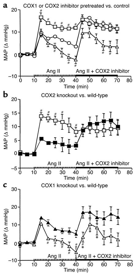
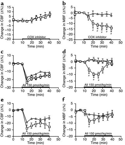
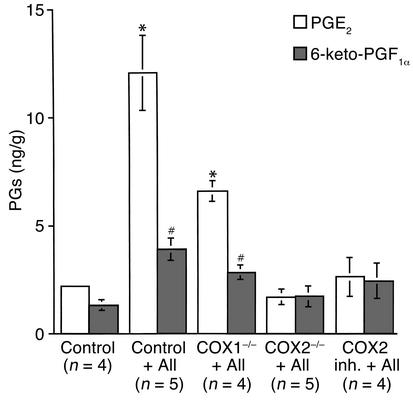
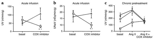
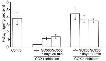
Therapeutic use of cyclooxygenase-inhibiting (COX-inhibiting) nonsteroidal antiinflammatory drugs (NSAIDs) is often complicated by renal side effects including hypertension and edema. The present studies were undertaken to elucidate the roles of COX1 and COX2 in regulating blood pressure and renal function. COX2 inhibitors or gene knockout dramatically augment the pressor effect of angiotensin II (Ang II). Unexpectedly, after a brief increase, the pressor effect of Ang II was abolished by COX1 deficiency (either inhibitor or knockout). Ang II infusion also reduced medullary blood flow in COX2-deficient but not in control or COX1-deficient animals, suggesting synthesis of COX2-dependent vasodilators in the renal medulla. Consistent with this, Ang II failed to stimulate renal medullary prostaglandin E(2) and prostaglandin I(2) production in COX2-deficient animals. Ang II infusion normally promotes natriuresis and diuresis, but COX2 deficiency blocked this effect. Thus, COX1 and COX2 exert opposite effects on systemic blood pressure and renal function. COX2 inhibitors reduce renal medullary blood flow, decrease urine flow, and enhance the pressor effect of Ang II. In contrast, the pressor effect of Ang II is blunted by COX1 inhibition. These results suggest that, rather than having similar cardiovascular effects, the activities of COX1 and COX2 are functionally antagonistic.


Figures

Figure 1
Figure 1
Breeding scheme for COX1 and COX2 knockout and wild-type mice used in these studies (see Methods for details).
Figure 2
Figure 2
Distinct effects of COX1 and COX2 on systemic arterial pressure. (a) The change in MAP (ΔMAP) following infusion of Ang II at 150 pmol/kg/min (between 10 and 40 minutes) and the addition of a COX2 inhibitor, SC58236 (between 40 and 70 minutes) in vehicle-treated mice (circles, n = 5); mice pretreated with SC58560, a COX1 inhibitor (triangles, n = 8); and mice pretreated with SC58236, the COX2 inhibitor (squares, n = 8). Ang II elevated MAP in vehicle-pretreated mice; the elevation was further increased upon addition of the COX2 inhibitor. Ang II increased significantly more in mice orally pretreated with the COX2 inhibitor, and significantly less in mice pretreated with the COX1 inhibitor (#P < 0.05 vs. control). (b) Ang II induced a higher increase of MAP in COX2 knockout mice (open squares, n = 6) than in wild-type controls (filled squares, n = 5; #P < 0.05). Additional COX2 inhibitor infusion further elevated MAP only in wild-type mice. (c) The pressor effect of Ang II was attenuated in COX1 knockout mice (open triangles, n = 6) versus wild-type controls (filled triangles, n = 5; #P < 0.05). MAP was further elevated by acute infusion of the COX2 inhibitor in both COX1 knockout and wild-type mice. *P < 0.05 vs. MAP at 40 minutes.
Figure 3
Figure 3
Effects of selective COX1 versus COX2 inhibition on cortical and medullary blood flow. (a and b) The effect of acute infusion of the COX1 inhibitor (triangles, n = 5) or COX2 inhibitor (squares, n = 4) on CBF and MBF in normal mice. *P < 0.05 vs. base-line MBF at 10 minutes; #P < 0.05 vs. MBF in the COX1 inhibitor–infused mice. (c and d) The effect of continuous Ang II infusion on CBF and MBF in mice pretreated with the COX2 inhibitor (squares, n = 7), COX1 inhibitor (triangles, n = 8), or vehicle (control group, circles, n = 5). *P < 0.05 vs. base-line MAP at 10 minutes; #P < 0.05 vs. control or COX1 inhibitor–treated mice. (e and f) The effects of Ang II infusion on CBF and MBF in COX2 knockout (squares, n = 6) and COX1 knockout mice (triangles, n = 6). *P < 0.05 vs. base-line MAP at 10 minutes; #P < 0.05 vs. COX1 knockout mice.
Figure 4
Figure 4
Distribution of COX1 and COX2 in mouse kidney. COX1 immunoreactivity in mouse outer medulla and papilla (a and b). COX2 immunoreactivity in cortex and medulla, respectively (c and d). Arrows indicate a vascular bundle in a and an intermediate-size arteriole in c.
Figure 5
Figure 5
Effect of Ang II infusion and COX1 and COX2 deficiency on renal medullary PGE2 and PGI2 synthesis. Prostanoids were measured by gas chromatography/mass spectrscopy in snap-frozen papillae dissected from mice pretreated with the COX2 inhibitor (COX2 inh.), vehicle-treated mice, or COX knockout mice with or without acute infusion of Ang II for 30 minutes. *P < 0.01 and #P < 0.05 vs. control.
Figure 6
Figure 6
(a and b) Effect of isozyme-selective COX inhibition on urinary volume (UV) (a) and sodium excretion (UNaV) (b). Urinary volume and sodium excretion were significantly reduced in mice receiving acute infusion of the COX2 inhibitor SC58236 (squares, n = 7) over 30 minutes, but not the COX1 inhibitor SC58560 (triangles, n = 5). *P < 0.05 vs. base-line; #P < 0.05 vs. the COX1 inhibitor. (c) The change in UV following acute Ang II infusion and sequential addition of Ang II plus a COX2 inhibitor in mice orally pretreated with the COX1 inhibitor (triangles, n = 12), COX2 inhibitor (squares, n = 8), and vehicle-treated controls (circles, n = 6). *P < 0.05 vs. base-line; #P < 0.05 vs. control or COX1 inhibitor–pretreated mice; P < 0.05 in controls and mice pretreated with COX1 inhibitor vs. UV stimulated by Ang II only.
Figure 7
Figure 7
PGE2 production in gastric mucosa from control, COX1-deficient, and COX2-deficient animals. From left to right, the bars represent PGE2 level in controls (n = 4), COX1 knockout mice (COX1–/–, n = 10), mice pretreated with the COX1 inhibitor for 7 days (n = 8), mice receiving acute infusion of the COX1 inhibitor for 30 minutes (n = 11), COX2 knockout mice (n = 7), mice pretreated with the COX2 inhibitor for 7 days (n = 8), and mice receiving acute infusion of the COX2 inhibitor for 30 minutes (n = 12). P < 0.05 in all three groups with COX1 inhibition vs. controls, or vs. each group with COX2 inhibition.

Comment in

References

    1. FitzGerald GA, Patrono C. The coxibs, selective inhibitors of cyclooxygenase-2. N Engl J Med. 2001;345:433–442. - PubMed
    1. Breyer MD, Harris RC. Cyclooxygenase 2 and the kidney. Curr Opin Nephrol Hypertens. 2001;10:89–98. - PubMed
    1. Whelton A, et al. Cyclooxygenase-2-specific inhibitors and cardiorenal function: a randomized, controlled trial of celecoxib and rofecoxib in older hypertensive osteoarthritis patients. Am J Ther. 2001;8:85–95. - PubMed
    1. Page J, Henry D. Consumption of NSAIDs and the development of congestive heart failure in elderly patients: an underrecognized public health problem. Arch Intern Med. 2000;160:777–784. - PubMed
    1. DeWitt DL, Smith WL. Primary structure of prostaglandin G/H synthase from sheep vesicular gland determined from the complementary DNA sequence. Proc Natl Acad Sci USA. 1988;85:1412–1416. - PMC - PubMed

Publication types

MeSH terms