. 2002 Jul;110(1):91-9.
doi: 10.1172/JCI13275.

G(1) and G(2) cell-cycle arrest following microtubule depolymerization in human breast cancer cells

Affiliations

G(1) and G(2) cell-cycle arrest following microtubule depolymerization in human breast cancer cells

April L Blajeski et al. J Clin Invest. 2002 Jul.

Abstract

Microtubule-depolymerizing agents are widely used to synchronize cells, screen for mitotic checkpoint defects, and treat cancer. The present study evaluated the effects of these agents on normal and malignant human breast cell lines. After treatment with 1 microM nocodazole, seven of ten breast cancer lines (type A cells) arrested in mitosis, whereas the other three (type B cells) did not. Similar effects were observed with 100 nM vincristine or colchicine. Among five normal mammary epithelial isolates, four exhibited type A behavior and one exhibited type B behavior. Further experiments revealed that the type B cells exhibited a biphasic dose-response curve, with mitotic arrest at low drug concentrations (100 nM nocodazole or 6 nM vincristine) that failed to depolymerize microtubules and a p53-independent p21(waf1/cip1)-associated G(1) and G(2) arrest at higher concentrations (1 microM nocodazole or 100 nM vincristine) that depolymerized microtubules. Collectively, these observations provide evidence for coupling of premitotic cell-cycle progression to microtubule integrity in some breast cancer cell lines (representing a possible "microtubule integrity checkpoint") and suggest a potential explanation for the recently reported failure of some cancer cell lines to undergo nocodazole-induced mitotic arrest despite intact mitotic checkpoint proteins.


Figures
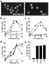
Figure 1
Figure 1
Two types of behavior following treatment of human breast cancer cell lines with microtubule-depolymerizing agents. (a) After a 24-hour treatment with 1 μM nocodazole, adherent and nonadherent MDA-MB-468 or MCF-7 cells were combined, stained with Hoechst 33258, and examined by fluorescence microscopy. Note that MDA-MB-468 cells remained in interphase, whereas many MCF-7 cells arrested in mitosis. (bd) Breast cancer cells (filled triangles, MCF-7; filled circles, HS0578T; open triangles, BT20; open circles, MDA-MB-468) were treated with 1 μM nocodazole (b), 100 nM vincristine (c), or 100 nM paclitaxel (d) for various periods of time. Alternatively, HMECs (e) were treated with these agents for 14 hours, a length of time chosen because this isolate doubled every 16 hours. After adherent and nonadherent cells were combined, the morphology of 300 or more nuclei in each sample was scored by fluorescence microscopy. In bd, representative individual experiments are shown and the degree of variation is indicated in the text. In e, the mean and range of two experiments are shown. Ctrl, control; Pacl, paclitaxel; Noco, nocodazole; Vin, vincristine.
Figure 2
Figure 2
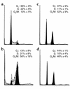
MDA-MB-468 cells arrest in G1 and G2 following nocodazole and vincristine treatment. Cells treated with diluent (a), 100 nM paclitaxel (b), 1 μM nocodazole (c), or 100 nM vincristine (d) for 24 hours were stained with PI and subjected to flow cytometry. Numbers in each panel show mean ± SD of distributions inferred from PI staining of three to six separate experiments using ModFit.
Figure 3
Figure 3
A subset of MDA-MB-468 cells arrest in late G2 following nocodazole or vincristine treatment. Cells growing on coverslips were treated with diluent (a and d), 1 μM nocodazole (b and e), or 100 nM vincristine (c and f) for 24 hours, then fixed and stained with antibodies to CENP-F (ac) or cyclin B (df). (g) The percentage of cells displaying focal nuclear CENP-F localization (arrow in a) increased after a 24-hour treatment with nocodazole or vincristine. Results shown are mean ± SD of three separate experiments. Ionizing radiation was used as a positive control. Quantitative analysis demonstrated that the number of cells with cytoplasmic cyclin B localization increased in a very similar fashion.
Figure 4
Figure 4
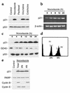
Increased p21 expression after treatment of MDA-MB-468 cells with microtubule-depolymerizing agents. (a) Cells were treated with 1 μM nocodazole, 100 nM paclitaxel, 100 nM vincristine, or 100 nM colchicine for 24 hours. Whole-cell lysates were subjected to SDS-PAGE and probed for p21. p53, which is mutated in these cells (41), served as a loading control. (b) Poly(A)+ RNA from cells treated with 1 μM nocodazole for various periods of time was analyzed by RT-PCR using primers specific for p21. β-actin served as a control. (c) Cells were treated with 1 μM nocodazole for various lengths of time before SDS-PAGE and immunoblotting for p21, GADD45 (GD45), or p53 (as a loading control). (d and e) Cells were treated with 1 μM nocodazole for 24 hours, fixed in 50% ethanol, stained with PI, and sorted into 2N and 4N populations by flow cytometry (gates set as shown). Protein (50 μg) from unsorted (total) or sorted (2N or 4N) cell populations was subjected to SDS-PAGE followed by immunoblotting. Cyclin B and cyclin E served as markers of the G2 and G1 populations, respectively, while PARP served as a loading control.
Figure 5
Figure 5
Dose-dependent mitotic arrest of type B cells during nocodazole treatment. (ad) After a 24-hour treatment with various nocodazole concentrations, adherent and nonadherent MDA-MB-468 (a), T47D (b), or MCF-7 cells (d) were combined and examined by fluorescence microscopy. (c) HMECs (passages 2–4) were treated for 24 hours with diluent (i), 100 nM paclitaxel (ii), or nocodazole at 83, 167, 1,200, and 3,000 nM (iii–vi, respectively). (e) After a 24-hour treatment with various concentrations of paclitaxel, adherent and nonadherent MDA-MB-468 cells were combined and examined by fluorescence microscopy. (fh) Whole-cell lysates from MDA-MB-468 cells treated with the indicated concentration of nocodazole for 24 hours (f), or the indicated cell lines treated with 1 μM nocodazole for 24 hours (g and h) were subjected to SDS-PAGE followed by blotting for p21 and PARP (which served as a loading control). Error bars show ± one SD from three independent experiments (a, b, d, and e), or ± range from two (c) independent experiments.
Figure 6
Figure 6
Effect of nocodazole on microtubule stability in MDA-MB-468 and MCF-7 cells. MDA-MB-468 (ac) or MCF-7 (df) cells growing on coverslips were treated with diluent (a and d), 167 nM nocodazole (b and e), or 1 μM nocodazole (c and f) for 8 hours (by this time most MCF-7 cells had not yet accumulated in mitosis and thus remained adherent), fixed, stained with anti–α-tubulin, and visualized by fluorescence microscopy.

References

    1. Jordan MA, Wilson L. Microtubules and actin filaments: dynamic targets for cancer chemotherapy. Curr Opin Cell Biol. 1998;10:123–130. - PubMed
    1. Downing KH. Structural basis for the interaction of tubulin with proteins and drugs that affect microtubule dynamics. Ann Rev Cell Dev Biol. 2000;16:89–111. - PubMed
    1. Rowinsky, E.K., and Donehower, R.C. 2001. Antimicrotubule agents. In Cancer chemotherapy and biotherapy. B.A. Chabner and D.L. Longo, editors. Lippincott, Williams & Wilkins. Philadelphia, Pennsylvania, USA. 329–372.
    1. Zieve GW, Turnbull D, Mullins JM, McIntosh JR. Production of large numbers of mitotic mammalian cells by use of the reversible microtubule inhibitor nocodazole. Exp Cell Res. 1980;126:397–405. - PubMed
    1. De Brabander M, Geuens G, Nuydens R, Willebrords R, De Mey J. Microtubule assembly in living cells after release from nocodazole block: the effects of metabolic inhibitors, taxol and pH. Cell Biol Int Rep. 1981;5:913–920. - PubMed

Publication types